. 2015 May 21;11(5):e1005134.
doi: 10.1371/journal.pgen.1005134. eCollection 2015 May.

Turning Escherichia coli into a Frataxin-Dependent Organism

Affiliations

Turning Escherichia coli into a Frataxin-Dependent Organism

Béatrice Roche et al. PLoS Genet. .

Abstract

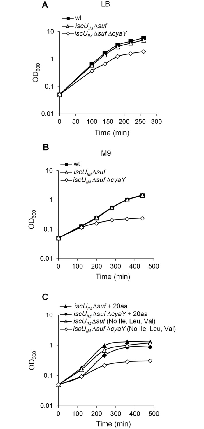
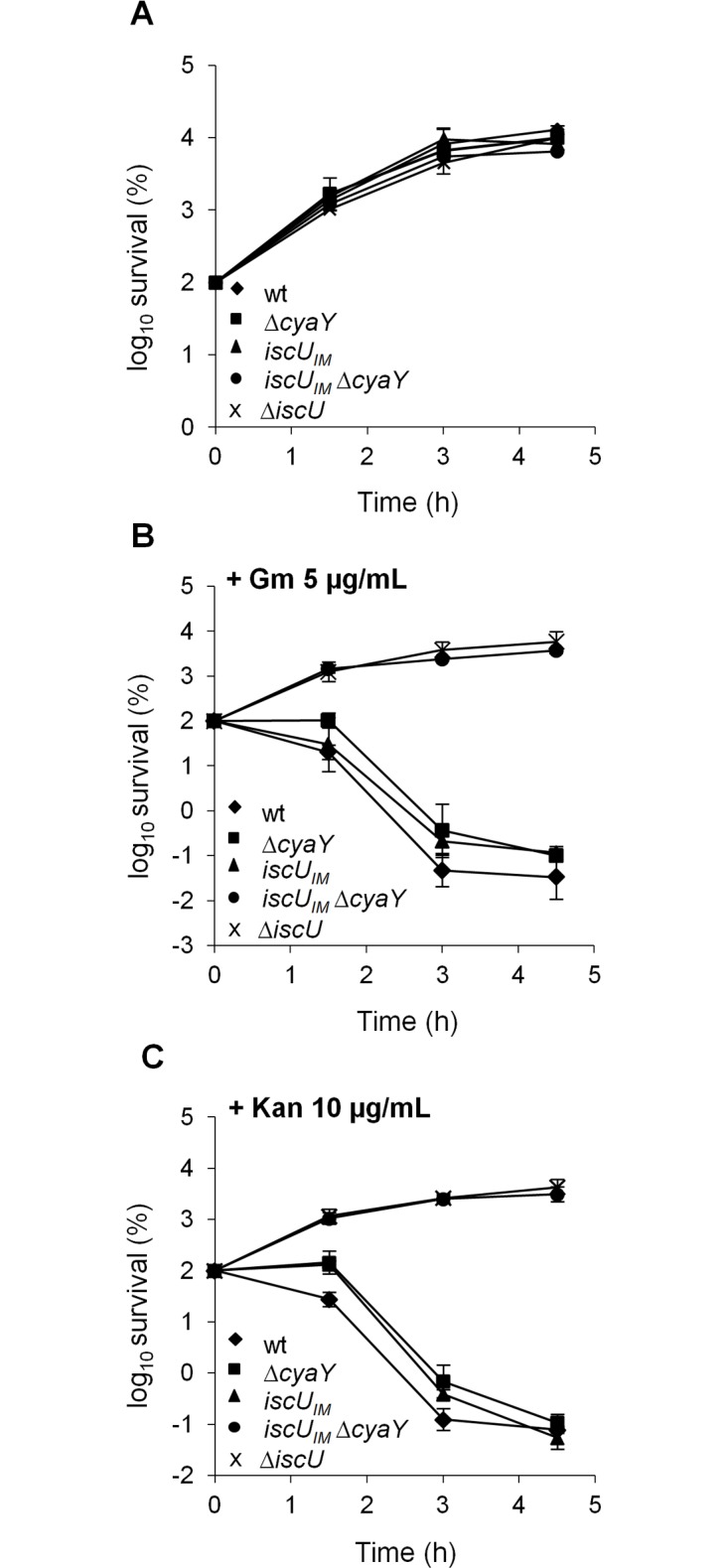
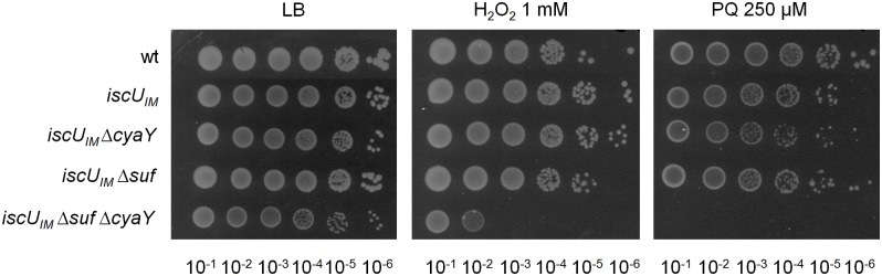
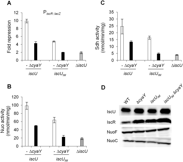
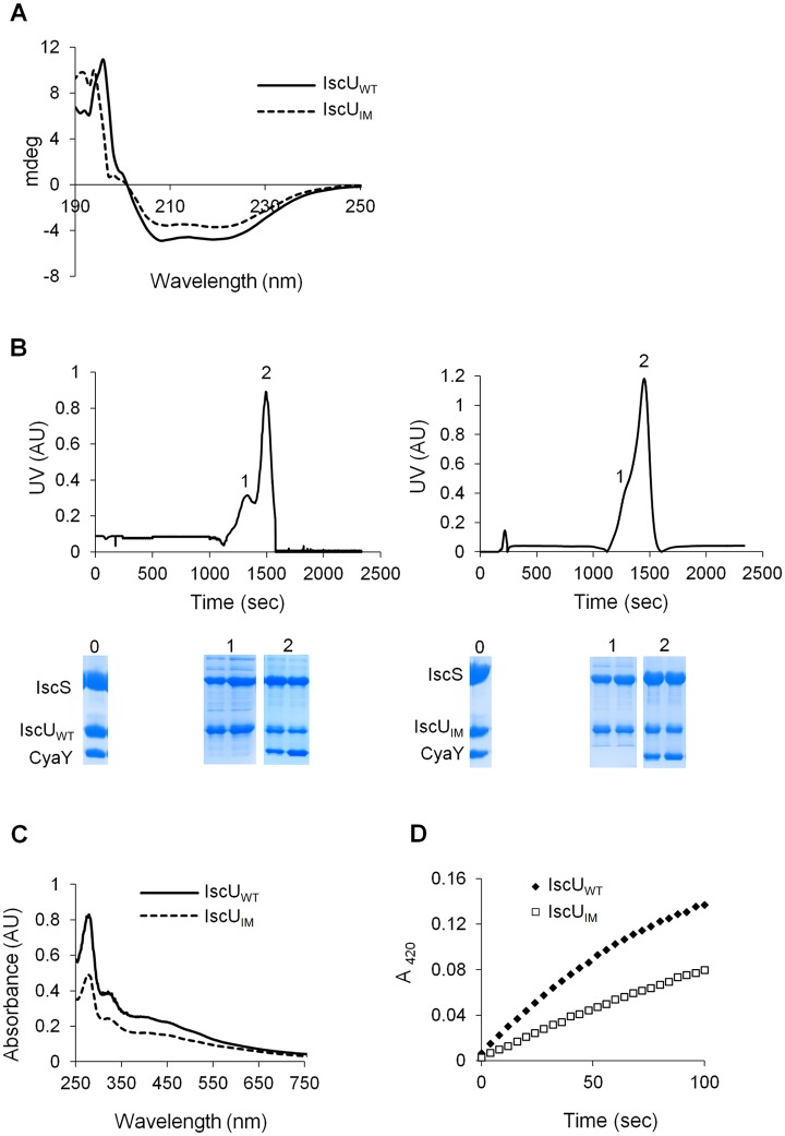
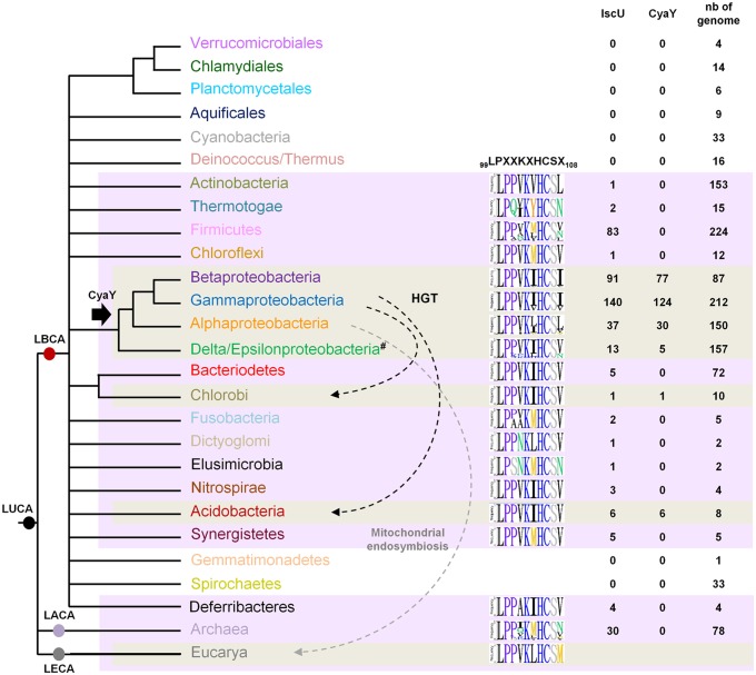
Fe-S bound proteins are ubiquitous and contribute to most basic cellular processes. A defect in the ISC components catalyzing Fe-S cluster biogenesis leads to drastic phenotypes in both eukaryotes and prokaryotes. In this context, the Frataxin protein (FXN) stands out as an exception. In eukaryotes, a defect in FXN results in severe defects in Fe-S cluster biogenesis, and in humans, this is associated with Friedreich's ataxia, a neurodegenerative disease. In contrast, prokaryotes deficient in the FXN homolog CyaY are fully viable, despite the clear involvement of CyaY in ISC-catalyzed Fe-S cluster formation. The molecular basis of the differing importance in the contribution of FXN remains enigmatic. Here, we have demonstrated that a single mutation in the scaffold protein IscU rendered E. coli viability strictly dependent upon a functional CyaY. Remarkably, this mutation changed an Ile residue, conserved in prokaryotes at position 108, into a Met residue, conserved in eukaryotes. We found that in the double mutant IscUIM ΔcyaY, the ISC pathway was completely abolished, becoming equivalent to the ΔiscU deletion strain and recapitulating the drastic phenotype caused by FXN deletion in eukaryotes. Biochemical analyses of the "eukaryotic-like" IscUIM scaffold revealed that it exhibited a reduced capacity to form Fe-S clusters. Finally, bioinformatic studies of prokaryotic IscU proteins allowed us to trace back the source of FXN-dependency as it occurs in present-day eukaryotes. We propose an evolutionary scenario in which the current mitochondrial Isu proteins originated from the IscUIM version present in the ancestor of the Rickettsiae. Subsequent acquisition of SUF, the second Fe-S cluster biogenesis system, in bacteria, was accompanied by diminished contribution of CyaY in prokaryotic Fe-S cluster biogenesis, and increased tolerance to change in the amino acid present at the 108th position of the scaffold.


Conflict of interest statement

The authors have declared that no competing interests exist.

Figures

Fig 1
Fig 1. The iscU IM Δsuf ΔcyaY strain exhibits growth defect.
Growth of wt (DV901), iscU IM Δ suf (BR763) and iscU IM Δ suf Δ cyaY (BR767) strains in LB (A). The wt (DV901), iscU IM Δ suf (BR763) and iscU IM Δ suf ΔcyaY (BR767) strains were grown overnight in glucose M9 minimal medium supplemented with all 20 amino acids. Cultures were then diluted into fresh glucose M9 minimal medium (B). Strains iscU IM Δ suf (BR763) and iscU IM Δ suf ΔcyaY (BR767) were grown overnight in glucose M9 minimal medium supplemented with all 20 amino acids. Cultures were then diluted into a fresh glucose M9 minimal medium supplemented with all amino acids or with all except Ile, Leu and Val (C). Growth was monitored at 600 nm. The experiment was repeated at least three times. One representative experiment is shown.
Fig 2
Fig 2. The iscU IM ΔcyaY strain is resistant to aminoglycosides.
Survival of wt (DV901), iscU IM (BR755) and their ΔcyaY derivatives (DV925 and BR756) without antibiotic (A) and after (B) Gentamicin (Gm) (5 μg/mL) and Kanamycin (Kan) (10 μg/mL) (C) treatment. Survival, measured by colony-forming units (CFU) per mL, was normalized relative to time zero at which the antibiotic was added (midexponential phase cells; ~5 ×10⁷ CFU/mL) and was plotted as log10 of % survival. Error bars represent the standard error from three independent experiments.
Fig 3
Fig 3. The iscU IM Δsuf ΔcyaY strain is hypersensitive to oxidative stress.
The wt (DV901), iscU IM (BR755), iscU IM ΔcyaY (BR756), iscU IM Δsuf (BR763) and iscU IM Δsuf ΔcyaY (BR767) strains were grown overnight at 37°C in LB medium. Cultures were diluted in sterile PBS, and 5 μL were directly spotted onto LB medium plates containing either 1 mM H2O2 or 250 μM paraquat. Growth was analysed after overnight incubation at 37°C. Each spot represents a 10-fold serial dilution.
Fig 4
Fig 4. Activities of Fe-S proteins in iscU IM and ΔcyaY strains.
Repression of the IscR-regulated gene (iscR::lacZ) (A), Nuo (B) and Sdh (C) activities in the wt (DV901) (white bars), iscU IM (BR755) (white bars), their ΔcyaY derivatives (DV925, BR756) (black bars), and ΔiscU (BR667) (grey bars) strains. The amount of IscR-dependent repression (fold repression) was determined by dividing the β-galactosidase activity present in the strain lacking IscR (DV915) by the β-galactosidase activity measured for each strain. Error bars represent the standard error from three independent experiments. (D) Cell extracts of indicated strains were subjected to immunoblot analysis using antibodies raised against IscU, IscR, NuoF and NuoC.
Fig 5
Fig 5. Analysis of IscUIM in vitro.
(A) Comparison of the CD spectra (expressed in mdeg) recorded in the region 190–250 nm between IscUWT (filled line) and IscUIM (dotted line). (B) Purified IscS, CyaY and IscUWT (left panel) or IscUIM (right panel) were mixed in 1:1:1 ratio (144 μM of each protein) in the presence of 4-fold excess of Fe(SO4)2(NH4)2, 10-fold excess of L-cysteine and 5 mM DTT and incubated for 40 minutes. The mixture was then loaded onto a QFF column equilibrated with 50 mM Tris pH 8. Proteins were eluted with 50 mM Tris pH 8 containing 1M NaCl. SDS-PAGE analyses have been performed on samples from the column on-put (0) and the peaks 1 and 2 for each mixture. (C) Reconstitution of [2Fe-2S] IscUWT (filled line) and IscUIM (dotted line) followed by UV-visible absorption spectroscopy. Apo-IscUWT or apo-IscUIM (144 μM) were incubated with 5 mM DTT, 1.44 μM IscS, 2 mM L-cysteine and 0.43 mM Fe(SO4)2(NH4)2 in 50 mM Tris-HCl pH 8. (D) Comparison of the kinetics of enzymatic Fe-S cluster formation on IscUWT (black diamonds) and IscUIM (white squares). Experiment was carried out using 25 μM IscUWT or IscUIM, 25 μM IscS, 100 μM Fe(SO4)2(NH4)2, 250 μM L-cysteine, 2 mM DTT. Fe-S cluster formation was followed by absorbance at 420 nm. The experiment was repeated at least three times. One representative experiment is shown.
Fig 6
Fig 6. Model for CyaY protein evolution.
Schematic representation of the universal tree of life, for which complete genome sequences are available. LUCA (Last Universal Common Ancestor), LECA (Last Eukaryotic Common Ancestor), LACA (Last Archaeal Common Ancestor) and LBCA (Last Bacterial Common Ancestor). For each prokaryotic phylum (whose color code is the same as the one used in S5 Fig), the number of genomes encoding a CyaY and a IscU homolog with respect to the number of complete available genomes is given. The black arrow indicates the presence of a CyaY encoding gene in the ancestor of a given lineage. The evolutionary event at the origin of the cyaY gene in the Delta/Epsilon subgroup cannot be definitively inferred. One hypothesis is that the cyaY gene is originated in the common ancestor of the Proteobacteria which together with a probable massive loss of cyaY (#) in Delta/Epsilonproteobacteria subgroup explains the presence of CyaY in the species of the Delta/Epsilonproteobacteria subgroup. Dotted arrows indicate horizontal gene transfer events (HGT) (black) and the mitochondrial endosymbiosis (grey). Sequence-logo of the region 99–108 in IscU homologs is also represented using Phylo-mLogo. This region contains the LPPVK motif and amino acid residues at position 108.

Comment in

References

    1. Py B, Barras F. Building Fe-S proteins: bacterial strategies. Nat Rev Microbiol. 2010;8: 436–446. 10.1038/nrmicro2356 - DOI - PubMed
    1. Crack JC, Green J, Thomson AJ, Le Brun NE. Iron-sulfur cluster sensor-regulators. Curr Opin Chem Biol. 2012;16: 35–44. 10.1016/j.cbpa.2012.02.009 - DOI - PubMed
    1. Balk J, Schaedler TA. Iron cofactor assembly in plants. Annu Rev Plant Biol. 2014;65: 125–53. 10.1146/annurev-arplant-050213-035759 - DOI - PubMed
    1. Roche B, Aussel L, Ezraty B, Mandin P, Py B, Barras F. Iron/sulfur proteins biogenesis in prokaryotes: formation, regulation and diversity. Biochim Biophys Acta. 2013;1827: 923–937. 10.1016/j.bbabio.2013.05.001 - DOI - PubMed
    1. Stehling O, Lill R. The role of mitochondria in cellular iron-sulfur protein biogenesis: mechanisms, connected processes, and diseases. Cold Spring Harb Perspect Med. 2013;3: 1–17. - PMC - PubMed

Publication types

LinkOut - more resources