. 2015 May 21;10(5):e0125698.
doi: 10.1371/journal.pone.0125698. eCollection 2015.

Persistent Foot-and-Mouth Disease Virus Infection in the Nasopharynx of Cattle; Tissue-Specific Distribution and Local Cytokine Expression

Affiliations

Persistent Foot-and-Mouth Disease Virus Infection in the Nasopharynx of Cattle; Tissue-Specific Distribution and Local Cytokine Expression

Juan M Pacheco et al. PLoS One. .

Abstract

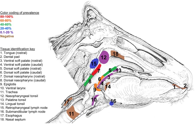
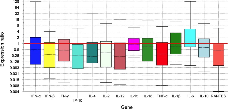
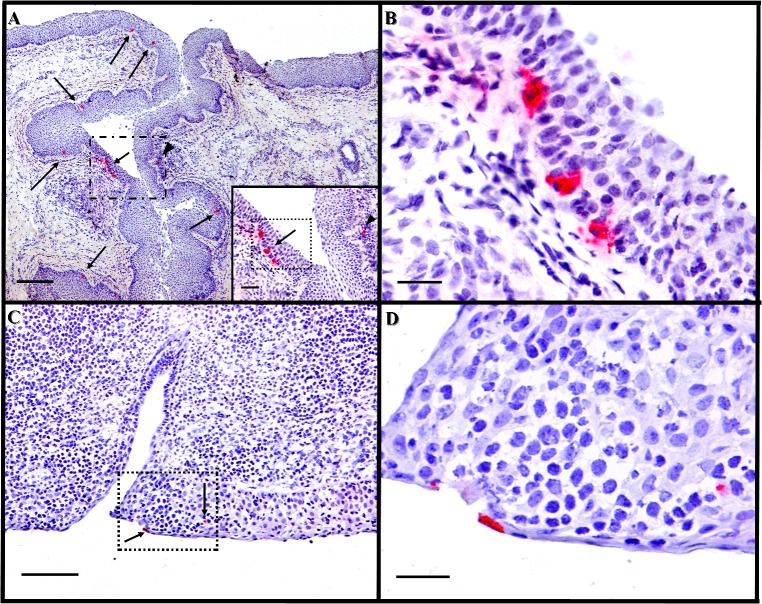
Tissues obtained post-mortem from cattle persistently infected with foot-and-mouth disease virus (FMDV) were analyzed to characterize the tissue-specific localization of FMDV and partial transcriptome profiles for selected immunoregulatory cytokines. Analysis of 28 distinct anatomic sites from 21 steers infected with FMDV serotype A, O or SAT2, had the highest prevalence of overall viral detection in the dorsal nasopharynx (80.95%) and dorsal soft palate (71.43%). FMDV was less frequently detected in laryngeal mucosal tissues, oropharyngeal mucosal sites, and lymph nodes draining the pharynx. Immunomicroscopy indicated that within persistently infected mucosal tissues, FMDV antigens were rarely detectable within few epithelial cells in regions of mucosa-associated lymphoid tissue (MALT). Transcriptome analysis of persistently infected pharyngeal tissues by qRT-PCR for 14 cytokine genes indicated a general trend of decreased mRNA levels compared to uninfected control animals. Although, statistically significant differences were not observed, greatest suppression of relative expression (RE) was identified for IP-10 (RE = 0.198), IFN-β (RE = 0.269), IL-12 (RE = 0.275), and IL-2 (RE = 0.312). Increased relative expression was detected for IL-6 (RE = 2.065). Overall, this data demonstrates that during the FMDV carrier state in cattle, viral persistence is associated with epithelial cells of the nasopharynx in the upper respiratory tract and decreased levels of mRNA for several immunoregulatory cytokines in the infected tissues.


Conflict of interest statement

Competing Interests: The authors have declared that no competing interests exist.

Figures

Fig 1
Fig 1. Tissue-specific distribution of FMDV.
Detection was performed by virus isolation or rRT-PCR in persistently infected steers inoculated by direct (intradermolingual route) or contact exposure inoculation. Only the epithelium of the Dorsal Nasopharynx (7) occupies the highest stratum of 80–100% indicating this tissue as the most consistent site of FMDV persistent infection. Prevalence values were calculated as number of animals in which a tissue was determined positive by one or both techniques/total number of animals.
Fig 2
Fig 2. Analysis of relative expression of different cytokines in dorsal soft palate of persistently infected steers.
Boxes represent the interquartile range, or the middle 50% of observations. The dotted line represents the median gene expression. Whiskers represent the minimum and maximum observations.
Fig 3
Fig 3. Immunohistochemical detection of persistent FMDV in nasopharyngeal mucosa.
A) Dorsal soft palate (caudal), steer #626, 37 dpe, FMDV O1 Manisa. FMDV non-structural protein localized to multiple cells within basal layers of crypt epithelium (arrows) as well as a single cell within associated lymphoid follicle (arrow head). 4x magnification, scale bar 200μm. Anti-FMDV 3D monoclonal antibody. Micropolymer alkaline phosphatase. Gill s hematoxylin counterstain. Inset; 20x magnification of region indicated in dashed rectangle in A, scale bar 50μm. B), 40x magnification of region within dashed rectangle in Fig 3A inset. Large, polygonal, immunopositive cells are within basal epithelium, scale bar 25 μm. C) Dorsal nasopharynx (rostral) steer #625, 37 dpi, FMDV O1 Manisa. FMDV structural protein localized to scarce individual cells within superficial and deeper layers of MALT-associated surface epithelium (arrows). 10x magnification, scale bar 100 μm. Anti-FMDV VP1 monoclonal antibody. Micropolymer alkaline phosphatase. Gill’s hematoxylin counterstain. D) 40x magnification of region within dashed rectangle in Fig 3C, superficial cells containing FMDV antigen are squamous, whereas deeper immunopositive cell is polygonal, scale bar 25 μm.
Fig 4
Fig 4. Immunofluorescent detection of persistent FMDV in nasopharyngeal mucosa.
Dorsal nasopharynx, steer #626, 37dpe, FMDV O1 Manisa. Immunomicroscopy, A) Low magnification (10X) view of an epithelial invagination with a focal cluster of FMDV-antigen-positive cells within superficial epithelial surface, scale bar 50 μm. B-F) Higher magnification (40X) views of region of interest identified in A (dashed box). Cells containing FMDV-VP1 are in the superficial epithelium and are cytokeratin-positive. Few MHC-II and CD11c positive cells are present within and below epithelium, but do not contain FMDV-VP1. Indirect fluorescence technique with differential interference contrast, antibody labels color-coded in bottom panel, asterisks indicate channels present in each panel, scale bar 25 μm.

Similar articles

Cited by

References

    1. Grubman MJ, Baxt B. Foot-and-mouth disease. Clin Microbiol Rev. 2004;17: 465–493. - PMC - PubMed
    1. OIE. Manual of Diagnostic Tests and Vaccines for Terrestrial Animals. 7th ed. Paris, France: World Organization for Animal Health; 2012. pp. 145–173.
    1. Alexandersen S, Zhang Z, Donaldson AI. Aspects of the persistence of foot-and-mouth disease virus in animals—the carrier problem. Microbes Infect. 2002;4: 1099–1110. - PubMed
    1. Arzt J, Juleff N, Zhang Z, Rodriguez LL. The pathogenesis of foot-and-mouth disease I: viral pathways in cattle. Transbound Emerg Dis. 2011;58: 291–304. 10.1111/j.1865-1682.2011.01204.x - DOI - PubMed
    1. Salt JS. Persistence of foot-and-mouth disease virus In: Sobrino F, Domingo E, editors. Foot and Mouth Disease Current Perspectives Wymondham, UK: Horizon Bioscience; 2004. pp. 103–143.

Publication types

MeSH terms

LinkOut - more resources