. 2015 May 21;10(5):e0127888.
doi: 10.1371/journal.pone.0127888. eCollection 2015.

DA-Raf-Mediated Suppression of the Ras--ERK Pathway Is Essential for TGF-β1-Induced Epithelial-Mesenchymal Transition in Alveolar Epithelial Type 2 Cells

Affiliations

DA-Raf-Mediated Suppression of the Ras--ERK Pathway Is Essential for TGF-β1-Induced Epithelial-Mesenchymal Transition in Alveolar Epithelial Type 2 Cells

Haruko Watanabe-Takano et al. PLoS One. .

Abstract

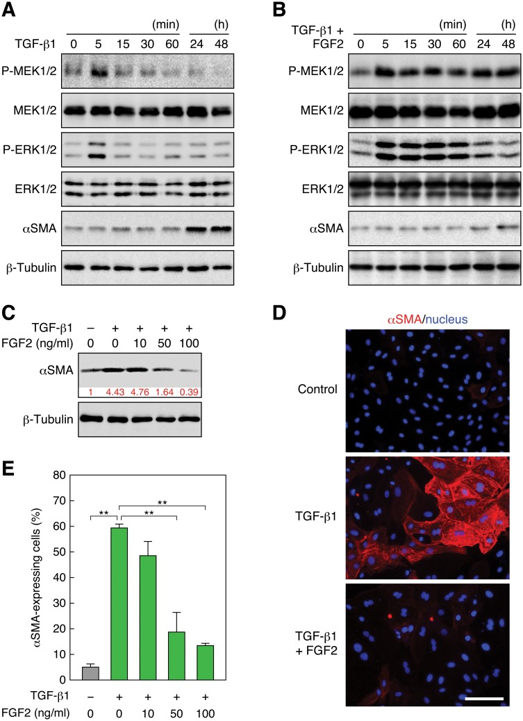
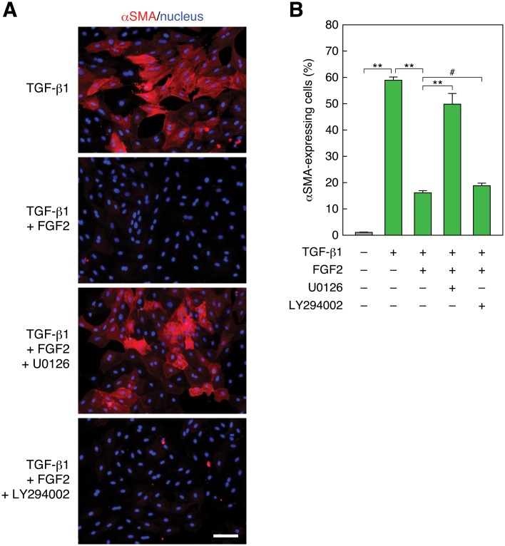
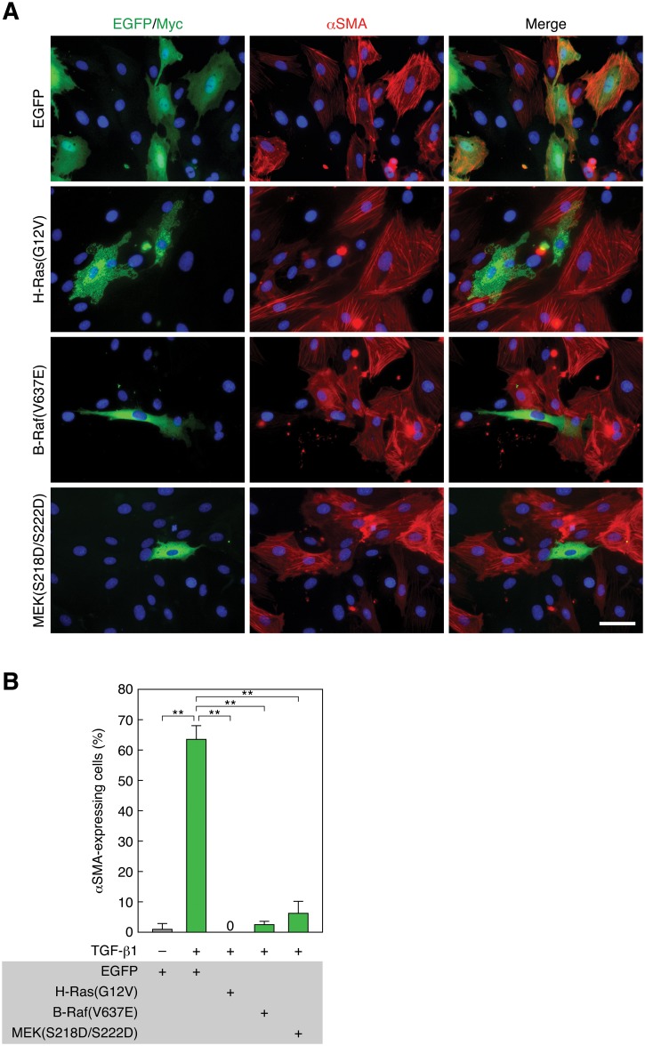
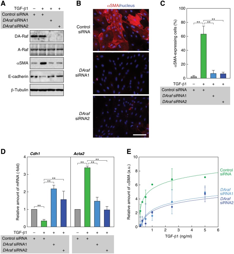
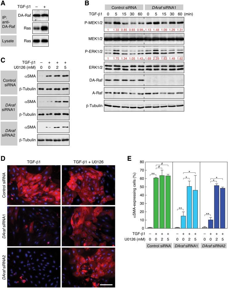
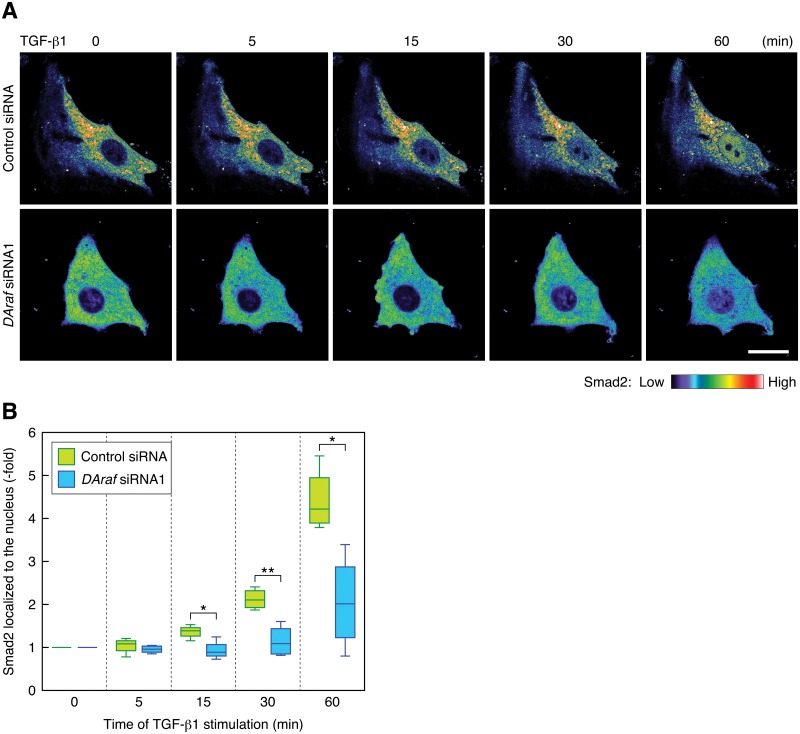
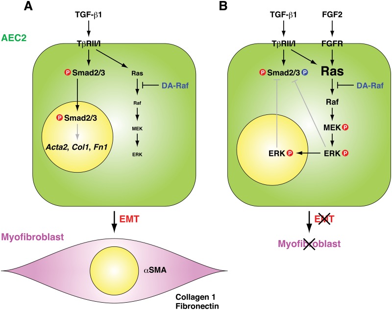
Myofibroblasts play critical roles in the development of idiopathic pulmonary fibrosis by depositing components of extracellular matrix. One source of lung myofibroblasts is thought to be alveolar epithelial type 2 cells that undergo epithelial-mesenchymal transition (EMT). Rat RLE-6TN alveolar epithelial type 2 cells treated with transforming growth factor-β1 (TGF-β1) are converted into myofibroblasts through EMT. TGF-β induces both canonical Smad signaling and non-canonical signaling, including the Ras-induced ERK pathway (Raf-MEK-ERK). However, the signaling mechanisms regulating TGF-β1-induced EMT are not fully understood. Here, we show that the Ras-ERK pathway negatively regulates TGF-β1-induced EMT in RLE-6TN cells and that DA-Raf1 (DA-Raf), a splicing isoform of A-Raf and a dominant-negative antagonist of the Ras-ERK pathway, plays an essential role in EMT. Stimulation of the cells with fibroblast growth factor 2 (FGF2), which activated the ERK pathway, prominently suppressed TGF-β1-induced EMT. An inhibitor of MEK, but not an inhibitor of phosphatidylinositol 3-kinase, rescued the TGF-β1-treated cells from the suppression of EMT by FGF2. Overexpression of a constitutively active mutant of a component of the Ras-ERK pathway, i.e., H-Ras, B-Raf, or MEK1, interfered with EMT. Knockdown of DA-Raf expression with siRNAs facilitated the activity of MEK and ERK, which were only weakly and transiently activated by TGF-β1. Although DA-Raf knockdown abrogated TGF-β1-induced EMT, the abrogation of EMT was reversed by the addition of the MEK inhibitor. Furthermore, DA-Raf knockdown impaired the TGF-β1-induced nuclear translocation of Smad2, which mediates the transcription required for EMT. These results imply that intrinsic DA-Raf exerts essential functions for EMT by antagonizing the TGF-β1-induced Ras-ERK pathway in RLE-6TN cells.


Conflict of interest statement

Competing Interests: The authors have declared that no competing interests exist.

Figures

Fig 1
Fig 1. FGF2 induces sustained activation of the Ras—ERK pathway and inhibits TGF-β1-induced EMT.
(A) Transient phosphorylation of MEK and ERK and induction of αSMA expression by TGF-β1 stimulation. RLE cells were stimulated with 0.5 ng/ml TGF-β1. The levels of MEK, phospho (P)-MEK, ERK, P-ERK, and αSMA, as well as β -tubulin as a standard, were analyzed by immunoblotting. (B) Sustained phosphorylation of MEK and ERK and inhibition of αSMA expression by FGF2 stimulation. RLE cells were stimulated with 100 ng/ml FGF2 in combination with 0.5 ng/ml TGF-β1. (C) A dose-dependent reduction of the TGF- β 1-induced αSMA protein level by FGF2 stimulation. RLE cells were stimulated with the indicated concentrations of FGF2 together with 0.5 ng/ml TGF- β 1 for 48 h. The relative intensity of αSMA band is indicated under the blot. (D) Induction of αSMA expression by TGF- β 1 and suppression of the expression by FGF2. RLE cells were stimulated with 0.5 ng/ml TGF- β 1 or with 100 ng/ml FGF2 along with TGF- β 1 for 48 h. αSMA expression and localization was detected by immunofluorescent staining with the Cy3—anti-αSMA mAb (red) as well as nuclear staining with Hoechst 33258 (blue). Scale bar, 50 μm. (E) A dose-dependent reduction of the ratio of TGF- β 1-induced αSMA-expressing cells by FGF2 stimulation. αSMA-expressing cells were detected as in (D). The values are means ± SD of 3 independent experiments. **, P < 0.01 by t test.
Fig 2
Fig 2. Inhibition of MEK but not PI3K recovers TGF-β1-induced and FGF2-suppressed EMT.
(A) Recovery of FGF2-suppressed αSMA expression by MEK inhibition but not by PI3K inhibition. RLE cells were pretreated with 10 μM of the MEK inhibitor U0126 or the PI3K inhibitor LY294002 for 30 min. Then they were stimulated with 0.5 ng/ml TGF-β1 along with 100 ng/ml FGF2 for 48 h. αSMA expression (red) and nuclei (blue) were detected as described in Fig 1 legend. Scale bar, 50 μm. (B) The ratio of αSMA-expressing cells in the analysis of (A). The values are means ± SD of 3 independent experiments. **, P < 0.01; #, P > 0.05 (not significant) by t test.
Fig 3
Fig 3. Constitutively active H-Ras, B-Raf, or MEK1 suppresses TGF-β1-induced EMT.
(A) Suppression of TGF-β1-induced EMT by constitutively active H-Ras, B-Raf, and MEK1. RLE cells were transfected with Myc-tagged H-Ras(G12V), EGFP-tagged B-Raf(V637E), EGFP—MEK1(S218D/S222D), or EGFP expression vector. Twenty-four hours after the transfection, they were treated with 0.5 ng/ml TGF-β1 for 48 h. αSMA expression (red) and nuclei (blue) were detected as described in Fig 1 legend. Myc- and EGFP-tagged proteins were detected by anti-Myc pAb and anti-GFP pAb staining, respectively (green). Scale bar, 20 μm. (B) The ratio of αSMA-expressing cells in the analysis of (A). The values are means ± SD of 3 independent experiments. **, P < 0.01 by t test.
Fig 4
Fig 4. Knockdown of DA-Raf abrogates TGF-β1-induced EMT.
(A) Suppression of TGF-β1-induced αSMA expression by knockdown of DA-Raf with DAraf siRNAs. RLE cells were transfected with DAraf siRNAs as well as a control siRNA. Twenty-four hours after the transfection, they were treated with 0.5 ng/ml TGF-β1 for 48 h. The levels of DA-Raf, A-Raf, αSMA, and E-cadherin, as well as β-tubulin as a standard, were analyzed by immunoblotting. (B) Suppression of TGF-β1-induced αSMA expression with DAraf siRNAs. RLE cells were transfected with DAraf siRNAs and treated with 0.5 ng/ml TGF-β1. αSMA expression (red) and nuclei (blue) were detected by fluorescence microscopy. Scale bar, 50 μm. (C) The ratio of αSMA-expressing cells in the analysis of (B). The values are means ± SD of 3 independent experiments. **, P < 0.01 by t test. (D) Elevation of the TGF-β1-suppressed Cdh1 (E-cadherin) mRNA level and suppression of the TGF-β1-induced Acta2 (αSMA) mRNA level with DAraf siRNAs. RLE cells were transfected with DAraf siRNAs and treated with TGF-β1 for 48 h. Relative levels of Cdh1 and Acta2 mRNAs normalized to the Actb (β-actin) mRNA level were determined by real-time PCR. The values are means ± SD of 3 independent experiments. **, P < 0.01 by t test. (E) Dose-dependent induction of αSMA expression by TGF-β1 and its suppression by DAraf siRNAs. RLE cells were transfected with DAraf siRNAs and treated with 0.1–5 ng/ml TGF-β1 for 48 h. The intensities of αSMA and β-tubulin bands on immunoblots were analyzed by densitometry. The graph shows the ratio of αSMA to β-tubulin band intensity against TGF-β1 concentration. The values are means ± SD of 3 independent experiments. a.u., arbitrary units.
Fig 5
Fig 5. Suppression of the ERK pathway by DA-Raf is required for TGF-β1-induced EMT.
(A) Induction of the binding of DA-Raf to Ras by TGF-β1 stimulation. The binding was analyzed by a coimmunoprecipitation assay. RLE cells were treated with 0.5 ng/ml TGF-β1 for 5 min. DA-Raf was immunoprecipitated with anti-DA-Raf pAb, and coprecipitated Ras was detected by immunoblotting with pan-Ras mAb. (B) Elevation of the phosphorylation levels of MEK and ERK by DA-Raf knockdown. RLE cells were transfected with DAraf siRNA1 as well as the control siRNA and treated with 0.5 ng/ml TGF-β1 for the indicated time. The levels of MEK, P-MEK, ERK, P-ERK, DA-Raf, A-Raf, and β-tubulin were analyzed by immunoblotting. The relative intensities of P-MEK1/2 and P-ERK1/2 bands are indicated under their blots. (C) Recovery of DAraf siRNA-blocked αSMA expression with U0126. RLE cells were transfected with DAraf siRNA1 or siRNA2 and then treated with 2 or 5 μM U0126 and 0.5 ng/ml TGF-β1 for 48 h. The level of αSMA, as well as β-tubulin as a standard, was analyzed by immunoblotting. (D) Recovery of DAraf siRNA-impaired αSMA expressing cells with U0126. RLE cells were transfected with DAraf siRNA1 or siRNA2 and then treated with 5 μM U0126 and 0.5 ng/ml TGF-β1 for 48 h. αSMA expression (red) and nuclei (blue) were detected. Scale bar, 50 μm. (E) The ratio of αSMA-expressing cells in the analysis of (D). The values are means ± SD of 3 independent experiments. *, P < 0.05; **, P < 0.01; #, P > 0.05 (not significant) by t test.
Fig 6
Fig 6. Knockdown of DA-Raf hinders the nuclear translocation of Smad2 induced by TGF-β1.
(A) Live cell images of the localization of Smad2. mCherry—Smad2-expressing RLE cells transfected with the control siRNA or DAraf siRNA1 were stimulated with 0.5 ng/ml TGF-β1 for the indicated time. The color indicator shows fluorescence intensity of mCherry—Smad2. Scale bar, 20 μm. (B) The degree of mCherry—Smad2 localization in the nucleus in the analysis of (A). Smad2 localized to the nucleus was calculated from the nuclear/cytoplasmic ratio of mCherry—Smad2 intensity. The box plot represents the data of 4 independent live cell images. *, P < 0.03; **, P < 0.005 by t test.
Fig 7
Fig 7. Postulated mechanisms of TGF-β1-induced EMT in RLE cells and the essential function of DA-Raf for EMT.
(A) TGF-β1-induced Smad2/3 signaling is essential for EMT from RLE cells to myofibroblasts expressing αSMA, collagen 1, and fibronectin. TGF-β1-induced Ras activity is weak, and intrinsic DA-Raf is sufficient to suppress the ERK pathway by binding to activated Ras. (B) FGF2 stimulation (as well as FGF1 or HGF stimulation) or overexpression of the constitutively active Ras, Raf, or MEK intensifies the activity of the Ras—ERK pathway activated by TGF-β1. Intrinsic DA-Raf is not sufficient to overcome the strong Ras—ERK pathway activity. Activated ERK might interfere with the nuclear translocation of Smad2/3, which is required to induce EMT.

Similar articles

Cited by

References

    1. Thannickal VJ, Toews GB, White ES, Lynch JP III, Martinez FJ (2004) Mechanisms of pulmonary fibrosis. Annu Rev Med 55: 395–417. - PubMed
    1. Selman M, Pardo A, Kaminski N (2008) Idiopathic pulmonary fibrosis: aberrant recapitulation of developmental programs? PLoS Med 5: e62 10.1371/journal.pmed.0050062 - DOI - PMC - PubMed
    1. King TE Jr, Pardo A, Selman M (2011) Idiopathic pulmonary fibrosis. Lancet 378: 1949–1961. 10.1016/S0140-6736(11)60052-4 - DOI - PubMed
    1. Willis BC, Liebler JM, Luby-Phelps K, Nicholson AG, Crandall ED, du Bois RM, et al. (2005) Induction of epithelial-mesenchymal transition in alveolar epithelial cells by transforming growth factor-β1: potential role in idiopathic pulmonary fibrosis. Am J Pathol 166: 1321–1332. - PMC - PubMed
    1. Kim KK, Kugler MC, Wolters PJ, Robillard L, Galvez MG, Brumwell AN, et al. (2006) Alveolar epithelial cell mesenchymal transition develops in vivo during pulmonary fibrosis and is regulated by the extracellular matrix. Proc Natl Acad Sci USA 103: 13180–13185. - PMC - PubMed

Publication types

MeSH terms

Substances