Vascular Parkinsonism: deconstructing a syndrome
- PMID: 25997420
- PMCID: PMC4478160
- DOI: 10.1002/mds.26263
Vascular Parkinsonism: deconstructing a syndrome
Abstract
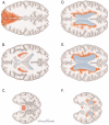
Progressive ambulatory impairment and abnormal white matter (WM) signal on neuroimaging come together under the diagnostic umbrella of vascular parkinsonism (VaP). A critical appraisal of the literature, however, suggests that (1) no abnormal structural imaging pattern is specific to VaP; (2) there is poor correlation between brain MRI hyperintensities and microangiopathic brain disease and parkinsonism from available clinicopathologic data; (3) pure parkinsonism from vascular injury ("definite" vascular parkinsonism) consistently results from ischemic or hemorrhagic strokes involving the SN and/or nigrostriatal pathway, but sparing the striatum itself, the cortex, and the intervening WM; and (4) many cases reported as VaP may represent pseudovascular parkinsonism (e.g., Parkinson's disease or another neurodegenerative parkinsonism, such as PSP with nonspecific neuroimaging signal abnormalities), vascular pseudoparkinsonism (e.g., akinetic mutism resulting from bilateral mesial frontal strokes or apathetic depression from bilateral striatal lacunar strokes), or pseudovascular pseudoparkinsonism (e.g., higher-level gait disorders, including normal-pressure hydrocephalus with transependimal exudate). These syndromic designations are preferable over VaP until pathology or validated biomarkers confirm the underlying nature and relevance of the leukoaraiosis. © 2015 International Parkinson and Movement Disorder Society.
Keywords: higher-level gait disorder; normal-pressure hydrocephalus; vascular parkinsonism; white matter ischemic disease.
© 2015 International Parkinson and Movement Disorder Society.
Figures


References
-
- Kumral E, Bayulkem G, Evyapan D, Yunten N. Spectrum of anterior cerebral artery territory infarction: Clinical and mri findings. Eur. J. Neurol. 2002;9:615–624. - PubMed
-
- Hama S, Yamashita H, Shigenobu M, Watanabe A, Kurisu K, Yamawaki S, et al. Post-stroke affective or apathetic depression and lesion location: Left frontal lobe and bilateral basal ganglia. Eur. Arch. Psychiatry Clin. Neurosci. 2007;257:149–152. - PubMed
-
- Critchley M. Arteriosclerotic parkinsonism. Brain. 1929;52:23–83.
-
- Thompson PD, Marsden CD. Gait disorder of subcortical arteriosclerotic encephalopathy: Binswanger's disease. Mov. Disord. 1987;2:1–8. - PubMed
-
- Mehta SH, Nichols FT, 3rd, Espay AJ, Duker AP, Morgan JC, Sethi KD. Dilated virchow-robin spaces and parkinsonism. Mov. Disord. 2013;28:589–590. - PubMed
Publication types
MeSH terms
Grants and funding
LinkOut - more resources
Full Text Sources
Other Literature Sources
Miscellaneous

