. 2015 May 22:6:7212.
doi: 10.1038/ncomms8212.

PI3K/mTORC2 regulates TGF-β/Activin signalling by modulating Smad2/3 activity via linker phosphorylation

Affiliations

PI3K/mTORC2 regulates TGF-β/Activin signalling by modulating Smad2/3 activity via linker phosphorylation

Jason S L Yu et al. Nat Commun. .

Abstract

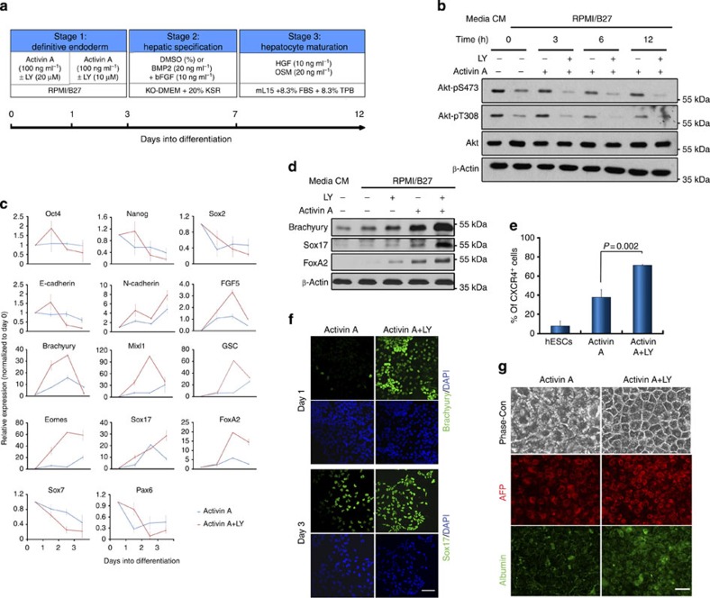
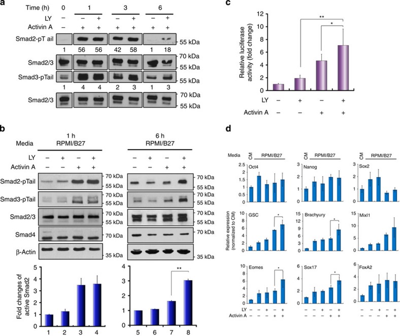
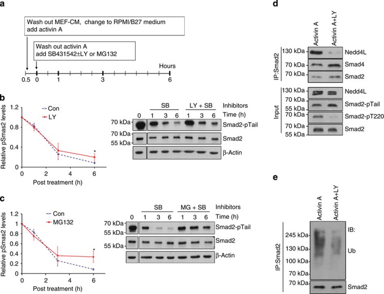
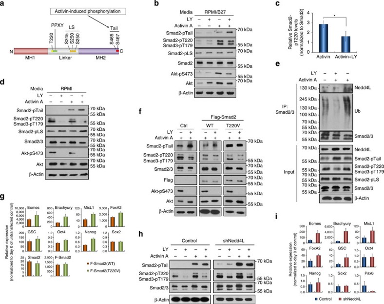
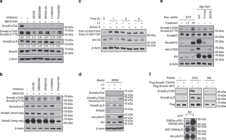
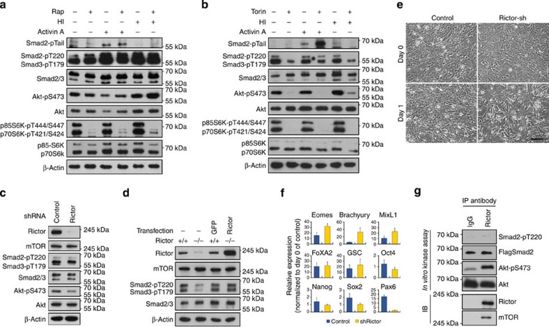
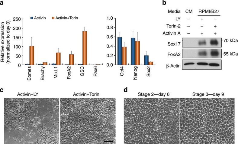
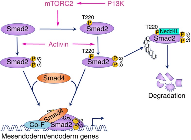
Crosstalk between the phosphatidylinositol 3-kinase (PI3K) and the transforming growth factor-β signalling pathways play an important role in regulating many cellular functions. However, the molecular mechanisms underpinning this crosstalk remain unclear. Here, we report that PI3K signalling antagonizes the Activin-induced definitive endoderm (DE) differentiation of human embryonic stem cells by attenuating the duration of Smad2/3 activation via the mechanistic target of rapamycin complex 2 (mTORC2). Activation of mTORC2 regulates the phosphorylation of the Smad2/3-T220/T179 linker residue independent of Akt, CDK and Erk activity. This phosphorylation primes receptor-activated Smad2/3 for recruitment of the E3 ubiquitin ligase Nedd4L, which in turn leads to their degradation. Inhibition of PI3K/mTORC2 reduces this phosphorylation and increases the duration of Smad2/3 activity, promoting a more robust mesendoderm and endoderm differentiation. These findings present a new and direct crosstalk mechanism between these two pathways in which mTORC2 functions as a novel and critical mediator.


Figures

Figure 1
Figure 1. Inhibition of PI3K signalling promotes differentiation of hESCs to the definitive endoderm (DE).
(a) Schematic illustrating the DE and hepatocyte differentiation protocol. (b) H1 hESCs cultured in MEF-CM (CM) were transferred into defined medium, RPMI/B27, for 1 h (time 0) prior to treatment with Activin A±LY294002 (LY). Cell lysates were collected at indicated time points and analysed by immunoblot. (c) Gene expression analysis by qRT–PCR in Activin A-treated hESCs with (red line) or without (blue line) LY. Data represent mean±s.d. from six measurements of two independent differentiations. (d) Immunoblot showing mesendoderm marker expression in hESCs treated for 24 h with indicated factors. (e) Percentage of CXCR4-positive cells by flow cytometry in hESCs with or without indicated treatment for 3 days. Data represent mean±s.d. from three independent biological samples. P value was calculated using the Student's t-test. (f) Immunostaining with Brachyury and Sox17 antibodies in hESCs treated as indicated. Scale bar, 50 μm. (g) hESCs were initially treated with Activin A±LY and then further differentiated to hepatocytes. Phase-contrast images (Phase-con) and immunostaining with AFP and albumin antibodies are presented. Scale bar, 50 μm.
Figure 2
Figure 2. Suppression of PI3K signalling prolongs Activin-induced Smad2/3 activity.
(a,b) hESCs were treated with Activin A or/and LY for the indicted time points before being subjected to immunoblotting analysis. Numbers in a represent the quantification of activated Smad2 and Smad3. Upper panels in b are representative immunoblot and lower panels are histograms of densitometric measurements from three independent biological samples. (c) Luciferase assay in hESCs co-transfected with pGL3-CAGA12-luc and renilla constructs and treated for 6 h. Data show mean±s.d. of three independent transfection experiments. (d) Messenger RNA expression of indicated markers by qRT–PCR. Bar graphs indicate fold induction. Data represent mean±s.d. of six measurements from two independent experiments. **P<0.001 and *P≤0.05 by the Student's t-test. LY, LY294002.
Figure 3
Figure 3. Inhibition of PI3K reduces Smad2/3 interaction with Nedd4L and subsequent ubiquitination.
(a) Scheme of treatment for b and c. (b,c) Representative immunoblot (right) and quantifications (left) of activated Smad2 (Smad2-pTail) in hESCs treated as illustrated in a with SB±LY (b) or SB±MG132 (c). Graphs represent mean±s.d. from three independent experiments. (d,e) hESCs treated with Activin A±LY for 1 h were analysed by Smad2 co-immunoprecipitation with indicated antibodies. *P≤0.05 by the Student's t-test. SB, SB431542; LY, LY294002; MG, MG132; Ub, ubiquitin.
Figure 4
Figure 4. PI3K regulates Nedd4L-mediated Smad2/3 degradation via phosphorylation of the T220/T179 linker residue.
(a) A schematic illustrating structural domains of Smad2 protein. The C-terminal and linker phosphorylation sites as well as the E3 ubiquitin ligase-binding PPXY motif are indicated. (b,c) Representative immunoblot (b) and quantification (c) showing significant reduction of Smad2-pT220 in hESCs at 6 h post treatment with Activin A+LY. Data in c represent mean±s.d. from three independent experiments. *P<0.05 (Student t-test). (d) Immunoblot showing that PC3 cells exhibited a similar pattern of Smad2 phosphorylation at various residues as that of hESCs in response to Activin±LY treatment after overnight starvation. (e) Nedd4L-mediated Smad2/3 ubiquitination requires phosphorylation at both linker T220/T179 and C-terminal SxS sites. PC3 cells were treated with Activin A±LY for 1 h, with MG132 added into the cultures 30 min prior to lysis. Smad2/3 immunoprecipitates were analysed by immunoblotting with indicated antibodies. (f) Immunoblot showing that Smad2-T220V mutant abolished the effect of LY on Activin-induced Smad2 activation. PC3 cells expressing Flag-tagged WT or T220V mutant (T220V) Smad2 were starved overnight, followed by treatment for 1 h with Activin A±LY. (g) qRT–PCR showing that hESCs expressing Smad2-T220V gave rise to higher levels of DE gene expression in response to Activin A. (h) Knockdown of Nedd4L in PC3 cells abolished the effect of LY on Activin-induced Smad2 activation. (i) qRT–PCR showing hESCs of Nedd4L knockdown exhibiting higher DE gene expression. hESCs in g and i were treated with Activin A for 2 days before the analysis, and data represent mean±s.d. from at least six measurements of two independent experiments.
Figure 5
Figure 5. Phosphorylation of Smad2/3 linker residues are differentially regulated by various signalling pathways.
(a,b) Immunoblot of Smad2/3 phosphorylation in hESCs that were pre-treated with Activin A, then incubated with indicated inhibitors for 1 h (a) or 6 h (b). (c) Immunoblot with Erk1/2 antibodies on cell extracts from hESCs with indicated treatments. (d) hESCs were starved for1 h in RPMI medium followed by treatment with either LY or HI for 1 h before being analysed by immunoblotting. (e) Hep3B cells transiently expressing either green fluorescent protein (GFP) or the various forms of Akt as indicated were starved overnight while GFP-expressing cells were treated with either LY or HI for 1 h before harvest for immunoblot. (f) In vitro kinase assay. Active Akt was isolated from PC3 cells by immunoprecipitation and incubated with recombinant GST-Flag-tagged wild-type (WT) or T220V mutant Smad2 in the presence of ATP. The protein mixtures were then resolved by SDS–polyacrylamide gel electrophoresis and analysed by immunoblotting with indicated antibodies. Commercially bought Erk2 and GST-GSK3 were used as positive controls. HI represents co-treatment with heregulin and IGF-1.
Figure 6
Figure 6. mTORC2 regulates Smad2/3 activity by modulating the phosphorylation of T220/T179 residue.
(a) Effect of rapamycin on Smad2/3 signalling. PC3 cells were treated with rapamycin in the presence of Activin A or HI for 6 h and then harvested for immunoblot with indicated antibodies. (b) Effect of Torin treatments on Activin-Smad2/3 signalling. Immunoblot of PC3 cells treated similar as in a but replacing rapamycin with Torin-2 (Torin). (c,d) Reduction of Smad2/3-pT220/T179 in rictor-shRNA knockdown PC3 cells (c) and in rictor null MEF (d). Ectopic expression of human rictor in rictor null MEF reverted the levels of Smad2/3-pT220/T179. (e,f) Knockdown of rictor in hESCs accelerated morphological changes associated with DE specification (e) and increased expression of DE markers (f) in response to Activin A. Scale bar, 100 μm. hESCs in f were treated with Activin A for 2 days before the analysis and data represent mean±s.d. from at least six measurements of two independent experiments. (g) In vitro kinase assay of mTORC2 on Smad2-T220 phosphorylation. mTORC2 complexes were isolated by immunoprecipitation with rictor antibody and incubated with recombinant GST-Flag-Smad2 or inactive Akt in the presence of ATP. The protein mixtures were then resolved by SDS–polyacrylamide gel electrophoresis and analysed by immunoblot.
Figure 7
Figure 7. Inhibition of mTOR enhances DE differentiation of hESCs.
(a) qRT–PCR showing gene expression in hESCs treated with Activin A±Torin for 2 days. Data represent mean±s.d. of six measurements from two independent experiments. (b) Immunoblot showing protein expression in hESCs treated as in a. (c) Phase-contrast images of hESCs treated for 3 days with Activin A together with either LY or Torin. Scale bar, 100 μm. (d) Phase-contrast images of the cells from c that were further differentiated by a hepatic differentiation protocol as shown in Fig. 1. Days of differentiation are indicated. Scale bar, 50 μm.
Figure 8
Figure 8. Model depicting a possible mechanism accounting for the inhibitory effect of PI3K/mTORC2 on the Activin-induced DE differentiation of hESCs.
.

References

    1. Massague J. & Wotton D. Transcriptional control by the TGF-beta/Smad signaling system. EMBO J. 19, 1745–1754 (2000). - PMC - PubMed
    1. Schmierer B. & Hill C. S. TGFbeta-SMAD signal transduction: molecular specificity and functional flexibility. Nat. Rev. Mol. Cell Biol. 8, 970–982 (2007). - PubMed
    1. Guo X. & Wang X. F. Signaling cross-talk between TGF-beta/BMP and other pathways. Cell Res. 19, 71–88 (2009). - PMC - PubMed
    1. Feng X. H. & Derynck R. Specificity and versatility in tgf-beta signaling through Smads. Annu. Rev. Cell Dev. Biol. 21, 659–693 (2005). - PubMed
    1. Massague J. TGFbeta signalling in context. Nat. Rev. Mol. Cell Biol. 13, 616–630 (2012). - PMC - PubMed

Publication types

MeSH terms

Substances