Review
. 2015 Dec:141:164-70.
doi: 10.1016/j.exer.2015.05.008. Epub 2015 May 18.

Electrophysiological assessment of retinal ganglion cell function

Affiliations
Review

Electrophysiological assessment of retinal ganglion cell function

Vittorio Porciatti. Exp Eye Res. 2015 Dec.

Abstract

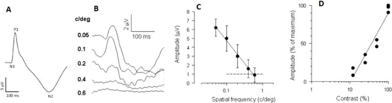
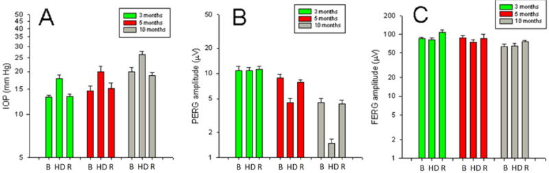
The function of retinal ganglion cells (RGCs) can be non-invasively assessed in experimental and genetic models of glaucoma by means of variants of the ERG technique that emphasize the activity of inner retina neurons. The best understood technique is the Pattern Electroretinogram (PERG) in response to contrast-reversing gratings or checkerboards, which selectively depends on the presence of functional RGCs. In glaucoma models, the PERG can be altered before histological loss of RGCs; PERG alterations may be either reversed with moderate IOP lowering or exacerbated with moderate IOP elevation. Under particular luminance-stimulus conditions, the Flash-ERG displays components that may reflect electrical activity originating in the proximal retina and be altered in some experimental glaucoma models (positive Scotopic Threshold response, pSTR; negative Scotopic Threshold Response, nSTR; Photopic Negative Response, PhNR; Oscillatory Potentials, OPs; multifocal ERG, mfERG). It is not yet known which of these components is most sensitive to glaucomatous damage. Electrophysiological assessment of RGC function appears to be a necessary outcome measure in experimental glaucoma models, which complements structural assessment and may even predict it. Neuroprotective strategies could be tested based on enhancement of baseline electrophysiological function that results in improved RGC survival. The use of electrophysiology in glaucoma models may be facilitated by specifically designed instruments that allow high throughput, robust assessment of electrophysiological function.

Keywords: Animal models; Electroretinogram; Glaucoma; Intraocular pressure; Pattern electroretinogram; Retinal ganglion cells.


Figures

Figure 1
Figure 1
Transient PERGs recorded in C57BL/6J mice in response to high-contrast gratings reversing 2 times/s. A: grand-average of 120 waveforms from different eyes.(Chou et al., 2014a) Note the N1-P1-N2 complex. B: The PERG amplitude decreases with increasing spatial frequency.(Porciatti, 2007) C: The mean (±SEM) PERG amplitude of 6 different mice decreases with increasing spatial frequency and reaches the noise level (dashed line) at 0.6 cy/deg (retinal acuity)(Porciatti, 2007), which is similar to the behavioral acuity.(Gianfranceschi et al., 1999) C: For gratings of 0.05 cy/deg, the PERG amplitude decreases with decreasing contrast. The contrast threshold is about 10%, (Porciatti et al., 1996) which is similar to that measured with the optomotor system. (Prusky et al., 2004)
Figure 2
Figure 2
Progressive loss of PERG amplitude in DBA/2J mice. A: serial PERG recordings in a population of DBA/2J mice (individual mice, grey lines). The PERG amplitude reaches the noise level (dashed line) at different ages between 8 and 12 months. Mice with PERG amplitude at noise level were eliminated from the pool for histological analysis. The open symbols connected by a thick line represent the group mean ± SEM amplitude. C: corresponding IOP measurements showing a progressive IOP increase with age. D: The PERG amplitude is inversely correlated to IOP. B: Relative changes of PERG amplitude (open circles), axon counts (open squares) and histological RNFL thickness (filled circles) as a function of age. Error bars represent the SD. Progressive loss of RGC axons and RNFL thickness lag behind progressive loss of PERG amplitude. Redrawn from ref. (Saleh et al., 2007). Axon counts estimates are from refs. (Anderson et al., 2005; Libby et al., 2005a)
Figure 3
Figure 3
Head-up posture reduces IOP and improves abnormal PERG in susceptible ages in DBA/2J mice. A: IOP decreases during 60° head-up posture (HU) by about 5 mm Hg compared to horizontal baseline (B) and recovery (R) in mice of different ages with different baseline IOP. B: PERG amplitude increases during HU in mice 6 and 10 months old, when baseline PERG is reduced. C: Improvement of PERG amplitude is also obtained in mice 11 months old with abnormal baseline PERG after mannitol 20% treatment, which reduced IOP by about 38%. Redrawn from ref.(Porciatti and Nagaraju, 2010)
Figure 4
Figure 4
Head-down posture increases IOP and reduces PERG in susceptible ages in DBA/2J mice. A: IOP increases during 60° head-down posture (HD) by about 5 mm Hg compared to horizontal baseline (B) and recovery (R) in mice of different ages with different baseline IOP. B: PERG amplitude decreases during HD in an age-dependent manner. C: The light-adapted, peak-to-trough flash ERG amplitude does not change during HD. Vertical Bars represent the mean, whiskers represent the SEM. Redrawn from ref. (Nagaraju et al., 2007)

References

    1. Abdul Y, Akhter N, Husain S. Delta-opioid agonist SNC-121 protects retinal ganglion cell function in a chronic ocular hypertensive rat model. Invest Ophthalmol Vis Sci. 2013;54:1816–1828. - PMC - PubMed
    1. Aihara M, Lindsey JD, Weinreb RN. Episcleral venous pressure of mouse eye and effect of body position. Curr Eye Res. 2003;27:355–362. - PubMed
    1. Anderson MG, Libby RT, Gould DB, Smith RS, John SW. High-dose radiation with bone marrow transfer prevents neurodegeneration in an inherited glaucoma. Proc Natl Acad Sci U S A. 2005;102:4566–4571. - PMC - PubMed
    1. Anderson MG, Libby RT, Mao M, Cosma IM, Wilson LA, Smith RS, John SW. Genetic context determines susceptibility to intraocular pressure elevation in a mouse pigmentary glaucoma. BMC Biol. 2006;4:20. - PMC - PubMed
    1. Bach M, Brigell MG, Hawlina M, Holder GE, Johnson MA, McCulloch DL, Meigen T, Viswanathan S. ISCEV standard for clinical pattern electroretinography (PERG): 2012 update. Doc Ophthalmol. 2013;126:1–7. - PubMed

Publication types