. 2015 Jul;36(7):777-85.
doi: 10.1017/ice.2015.73. Epub 2015 May 8.

Whole-genome sequencing for outbreak investigations of methicillin-resistant Staphylococcus aureus in the neonatal intensive care unit: time for routine practice?

Affiliations

Whole-genome sequencing for outbreak investigations of methicillin-resistant Staphylococcus aureus in the neonatal intensive care unit: time for routine practice?

Taj Azarian et al. Infect Control Hosp Epidemiol. 2015 Jul.

Abstract

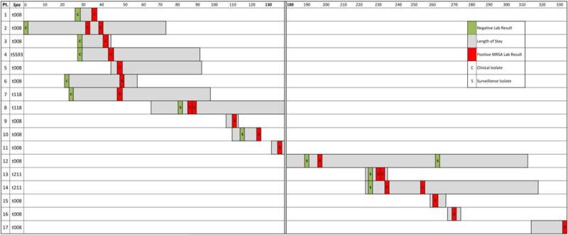
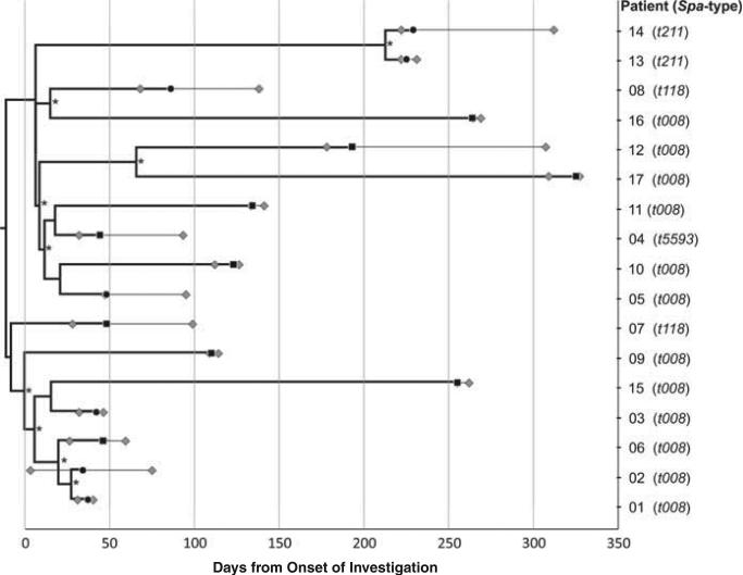
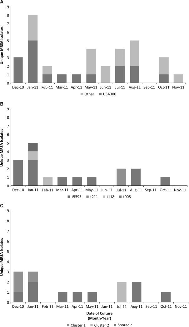
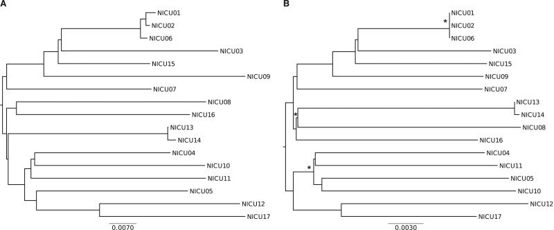
BACKGROUND Infants in the neonatal intensive care unit (NICU) are at increased risk for methicillin-resistant Staphylococcus aureus (MRSA) acquisition. Outbreaks may be difficult to identify due in part to limitations in current molecular genotyping available in clinical practice. Comparison of genome-wide single nucleotide polymorphisms (SNPs) may identify epidemiologically distinct isolates among a population sample that appears homogenous when evaluated using conventional typing methods. OBJECTIVE To investigate a putative MRSA outbreak in a NICU utilizing whole-genome sequencing and phylogenetic analysis to identify recent transmission events. DESIGN Clinical and surveillance specimens collected during clinical care and outbreak investigation. PATIENTS A total of 17 neonates hospitalized in a 43-bed level III NICU in northeastern Florida from December 2010 to October 2011 were included in this study. METHODS We assessed epidemiological data in conjunction with 4 typing methods: antibiograms, PFGE, spa types, and phylogenetic analysis of genome-wide SNPs. RESULTS Among the 17 type USA300 isolates, 4 different spa types were identified using pulsed-field gel electrophoresis. Phylogenetic analysis identified 5 infants as belonging to 2 clusters of epidemiologically linked cases and excluded 10 unlinked cases from putative transmission events. The availability of these results during the initial investigation would have improved infection control interventions. CONCLUSION Whole-genome sequencing and phylogenetic analysis are invaluable tools for epidemic investigation; they identify transmission events and exclude cases mistakenly implicated by traditional typing methods. When routinely applied to surveillance and investigation in the clinical setting, this approach may provide actionable intelligence for measured, appropriate, and effective interventions.


Figures

FIGURE 1
FIGURE 1
Detailed timeline for putative neonatal intensive care unit (NICU) outbreak. Lengths of stay are indicated in grey for the 17 pulse-field gel electrophoresis type USA300 isolates. Spa-types are indicated next to the patient number. Positive (red) and previously negative (green) surveillance (S) and clinical (C) isolates are illustrated. A 37-day gap between the 2 discrete outbreak periods is designated with a double vertical line shaded in gray.
FIGURE 2
FIGURE 2
Integration of epidemiological and phylogenetic data to produce a timeline of putative neonatal intensive care unit (NICU) outbreak. The Bayesian maximum clade credibility (MCC) tree represents the phylogenetic relationship between pulsed-field gel electrophoresis (PFGE)-type USA300 isolates from 17 infants hospitalized in the NICU. The MCC phylogeny is scaled in time with tip dates, indicated by black a circle (clinical isolates) or square (surveillance isolate), corresponding to the date of incident laboratory result. The lengths of stay of each infant are represented as lines extending from the phylogeny tip dates. The first gray diamond represents the day of admission scaled by the earliest admission date among all cases. The last gray diamond represents the date of discharge. An asterisk on the branch marks subtending clades supported by posterior probability >0.75. The Bayesian MCC tree of 17 NICU isolates was constructed using HKY nucleotide substitution model, Bayesian Skyline demographic model, and lognormal uncorrelated (relaxed) molecular clock.
FIGURE 3
FIGURE 3
Epidemic curve of putative (neonatal intensive care unit) NICU outbreak incorporating increasing levels of resolution from molecular and whole-genome sequencing (WGS) analysis. (A) Epidemic curve of 34 cases using dates of incident clinical or surveillance MRSA-positive laboratory results. Cases identified as USA300 using pulsed-field gel electrophoresis (PFGE) (n = 17) during the primary outbreak investigation are indicated in blue. (B) Epidemic curve of 17 PFGE-typed USA300 cases stratified by spa-type conducted retrospectively to identify 5 non-t008 spa-types among the 17 PFGE-typed USA300 isolates. (C) Epidemic curve of 10 remaining PFGE-typed USA300 and spa-type t008 isolates further stratified by results from phylogenetic analyses. Cluster 1 (patients 1, 2, and 6) represent epidemiological linkages based on phylogenetic data (eg, SNP distances) and epidemiological assessment (eg, overlapping lengths of stay).
FIGURE 4
FIGURE 4
Comparison of neighbor joining (NJ) trees constructed with genome wide single-nucleotide polymorphism data (A) and MLST + allelic profiles (B). Scale bars represent genetic distances based on nucleotide substitutions per SNP site (A) and allelic distance (B). MEGA v6.0 was used to infer phylogeny A using Kimura 2-parameter nucleotide substitution model, and branching patterns were evaluated by bootstrapping (1,000 replicates). Ridom's SeqSphere software package was used to create phylogeny B using MLST+ data from assembled genomes. Differences in the clustering of isolates are indicated with an asterisk. While branch lengths and genetic distance scale vary between phylogenies, the overall topology and interpretation remain largely unchanged.

References

    1. Gerber SI, Jones RC, Scott MV, et al. Management of outbreaks of methicillin-resistant Staphylococcus aureus infection in the neonatal intensive care unit: a consensus statement. Infect Control Hosp Epidemiol. 2006;27:139–145. - PubMed
    1. Saiman L, Cronquist A, Wu F. An outbreak of methicillin-resistant Staphylococcus aureus in a neonatal intensive care unit. Infect Control Hosp Epidemiol. 2003;24:317–321. - PubMed
    1. Geva A, Wright SB, Baldini LM, Smallcomb JA, Safran C, Gray JE. Spread of methicillin-resistant Staphylococcus aureus in a large tertiary NICU: network analysis. Pediatrics. 2011;128:e1173–e1180. - PMC - PubMed
    1. Iwamoto M, Mu Y, Lynfield R, et al. Trends in invasive methicillin-resistant Staphylococcus aureus infections. Pediatrics. 2013;132:e817–e824. - PMC - PubMed
    1. Zervou FN, Zacharioudakis IM, Ziakas PD, Mylonakis E. MRSA colonization and risk of infection in the neonatal and pediatric ICU: a meta-analysis. Pediatrics. 2014;133:e1015–e1023. - PubMed

Publication types

MeSH terms