Review
. 2015 Aug;16(5):320-30.
doi: 10.1111/pedi.12286. Epub 2015 May 22.

Organ donor specimens: What can they tell us about type 1 diabetes?

Affiliations
Review

Organ donor specimens: What can they tell us about type 1 diabetes?

Martha Campbell-Thompson. Pediatr Diabetes. 2015 Aug.

Abstract

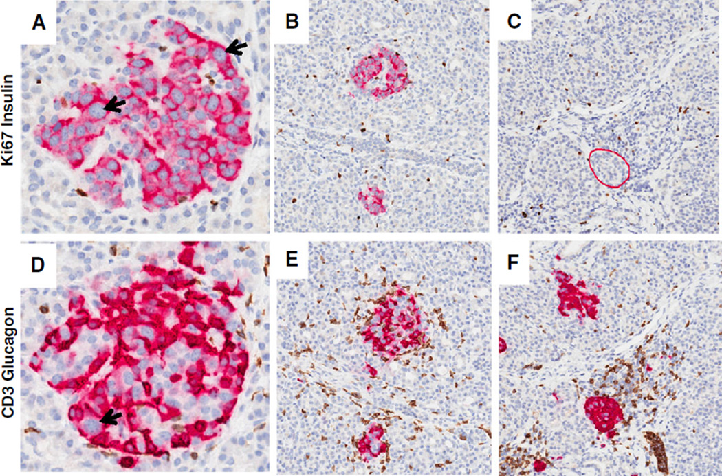
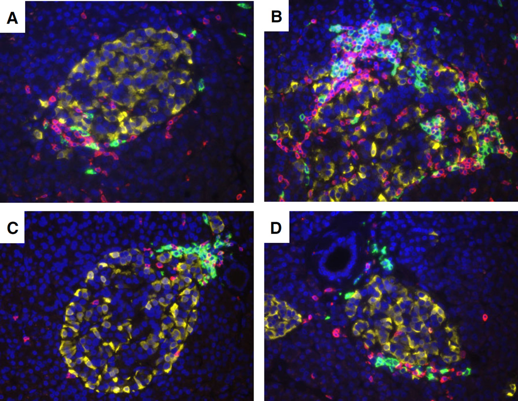
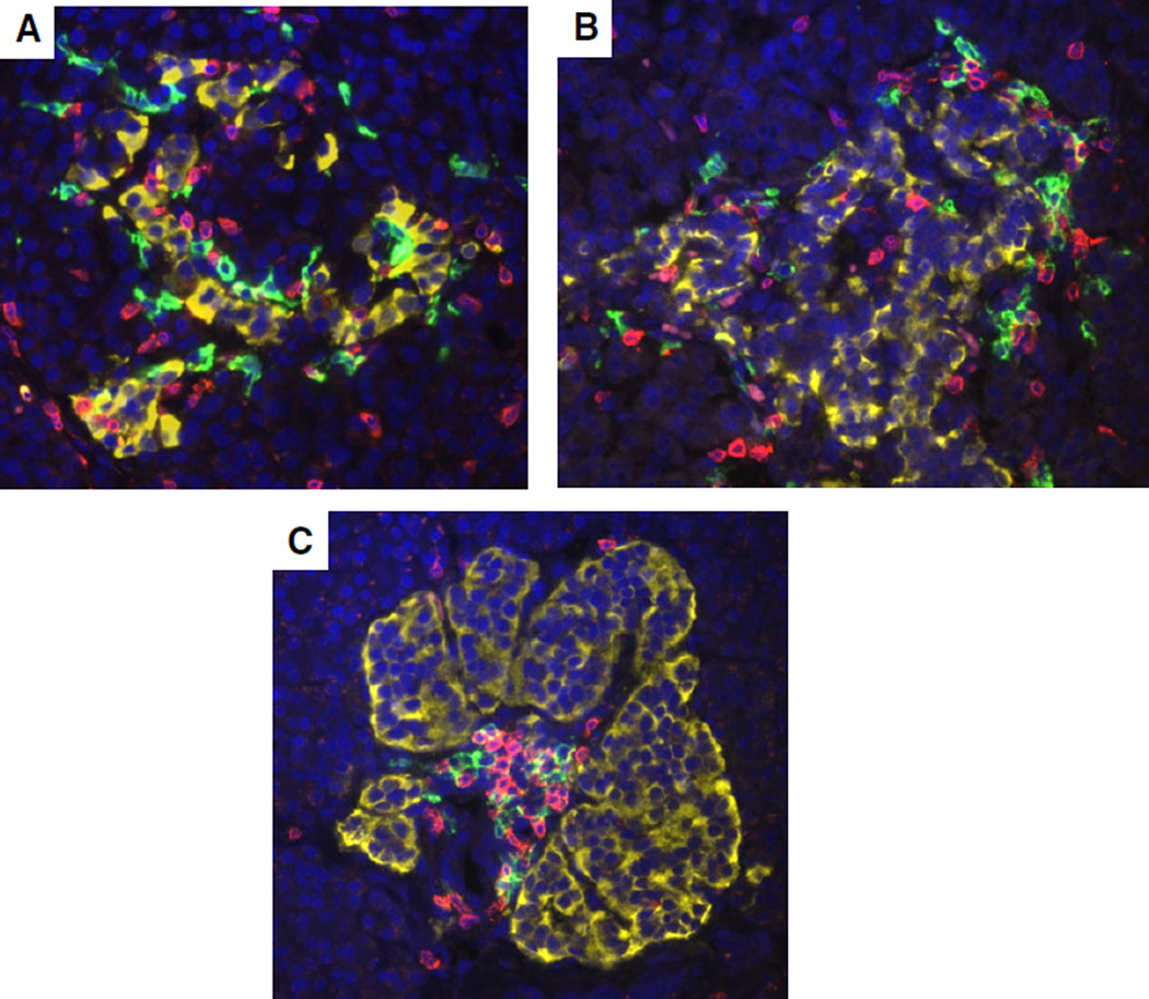
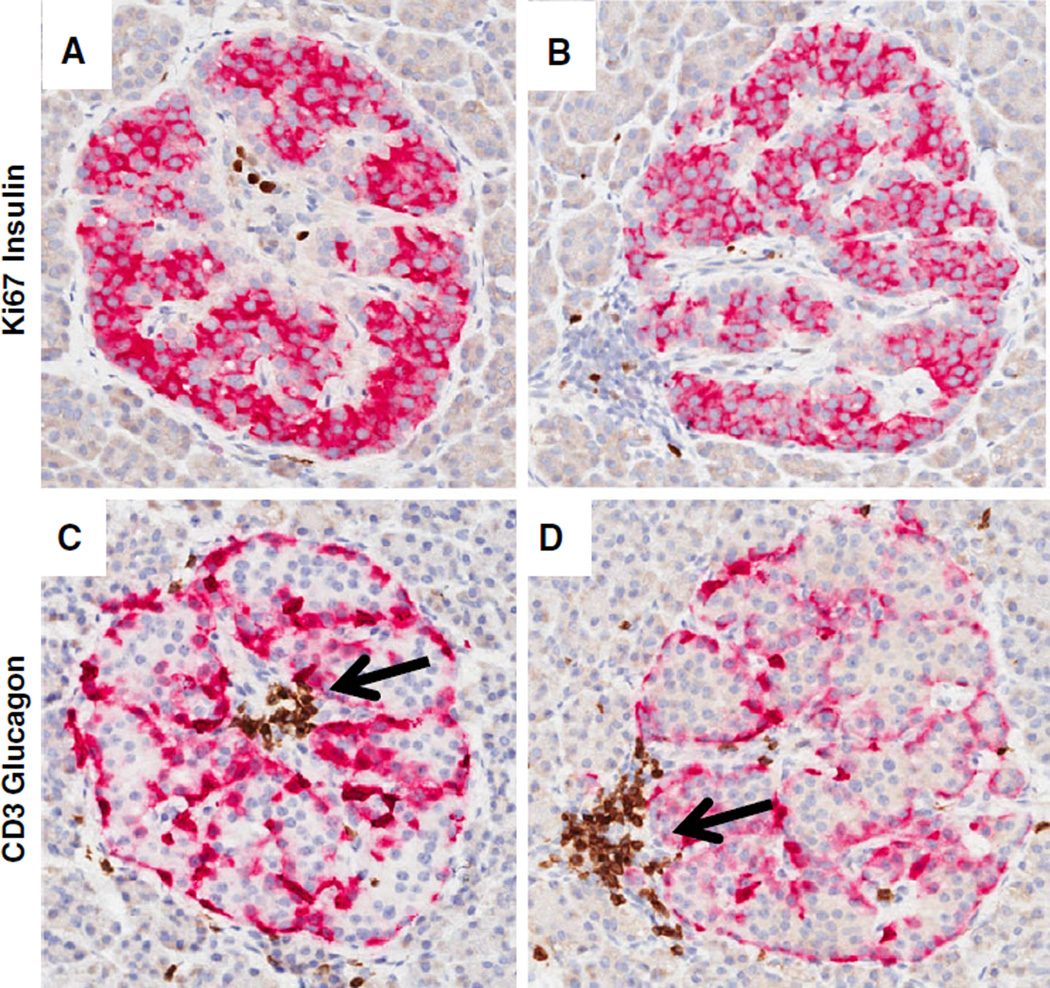
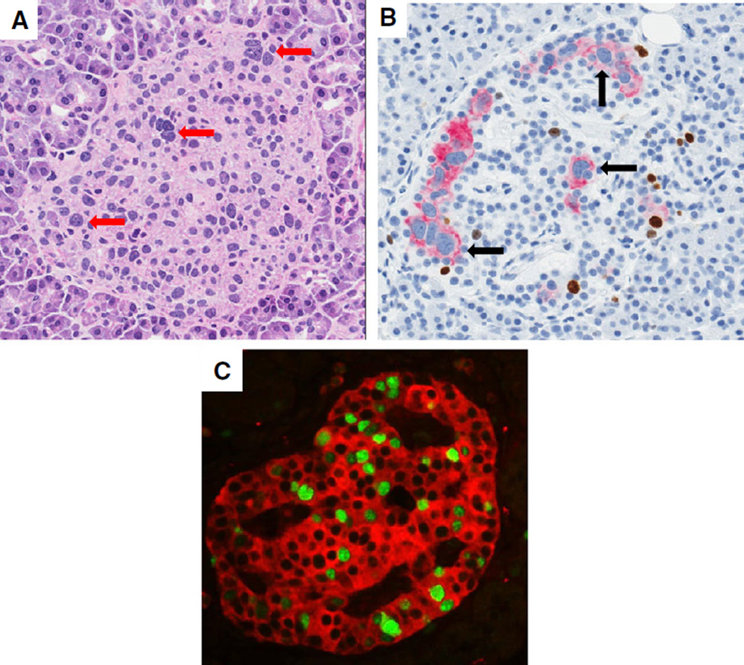
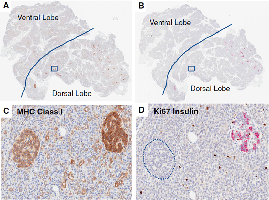
Type 1 diabetes (T1D) is a chronic disease resulting from the destruction of pancreatic beta cells, due to a poorly understood combination of genetic, environmental, and immune factors. The JDRF Network for Pancreatic Organ donors with Diabetes (nPOD) program recovers transplantation quality pancreas from organ donors throughout the USA. In addition to recovery of donors with T1D, non-diabetic donors include those with islet autoantibodies. Donors with type 2 diabetes and other conditions are also recovered to aid investigations directed at the full spectrum of pathophysiological mechanisms affecting beta cells. One central processing laboratory conducts standardized procedures for sample processing, storage, and distribution, intended for current and future cutting edge investigations. Baseline histology characterizations are performed on the pancreatic samples, with images of the staining results provided though whole-slide digital scans. Uniquely, these high-grade biospecimens are provided without expense to investigators, working worldwide, seeking methods for disease prevention and reversal strategies. Collaborative working groups are highly encouraged, bringing together multiple investigators with different expertise to foster collaborations in several areas of critical need. This mini-review will provide some key histopathological findings emanating from the nPOD collection, including the heterogeneity of beta cell loss and islet inflammation (insulitis), beta cell mass, insulin-producing beta cells in chronic T1D, and pancreas weight reductions at disease onset. Analysis of variations in histopathology observed from these organ donors could provide for mechanistic differences related to etiological agents and serve an important function in terms of identifying the heterogeneity of T1D.

Keywords: beta cell; image analysis; insulin; insulitis; islet.


Conflict of interest statement

Conflict of interest

The author declares no conflict of interest.

Figures

Fig. 1
Fig. 1
Insulitic islets at onset of T1D. Representative images of insulitic islets are shown from a 13-yr-old male Caucasian donor at T1D onset (GADA+ IA-2A+ ZnT8+; DR3/4; nPOD 6228). Serial paraffin sections were stained by two double immunohistochemistry stains (Ki67 and insulin, CD3 and glucagon) (17). Insulin + islets are shown in panels (A and B) and insulin-islets are shown in panel (C) (insulitic islet is circled in red). The islets in panels (D–F) show insulitis with varying numbers of CD3+ cells. CD3+ cells were found at the islet periphery and interior region. Panels (E) and (F) also display examples of the large numbers of CD3+ cells in the exocrine region. Islet endocrine cell nuclear pleomorphism is also shown in panels (A) and (D) (arrows). Additional donor information and whole slide images are available through the nPOD Online Pathology Database (http://www.jdrfnpod.org/for-investigators/online-pathology-information/). Original magnification: 20×. GADA, glutamic acid decarboxylase-65 autoantibody; IA-2A, islet antigen-2 or insulinoma antigen-2; nPOD, Network for Pancreatic Organ donors with Diabetes; T1D, type 1 diabetes; ZnT8, zinc transporter 8 autoantibody.
Fig. 2
Fig. 2
CD20+ and CD3+ lymphocyte numbers between insulitic islets in a young donor with recent onset. Representative images of four islets with insulitis are shown from a 12-yr-old male African American donor 1 yr after onset (IA-2A+, DR09/16 nPOD 6052). The paraffin section was stained by multiple immunofluorescence for CD20 (green), CD3 (yellow), and glucagon (red) using methods previously described (17). Total numbers of CD20+ and CD3+ cells varied by islet and their locations were at the islet periphery (A–D) and interior (B) Original magnification: 40×. IA-2A, islet antigen-2 or insulinoma antigen-2; nPOD, Network for Pancreatic Organ donors with Diabetes.
Fig. 3
Fig. 3
CD20+ and CD3+ lymphocytes in insulitic islets with varying disease durations. Representative islets are shown from a 5-yr-old female Caucasian donor with T1D for 3 months (IA-2A+ ZnT8A+, DR3/4, nPOD 6209, A), a 13-yr-old male Caucasian donor at onset (GADA+ IA-2A+ ZnT8+; DR3/4, nPOD 6228, B), and a 19-yr-old male Caucasian donor with T1D for 5 yr (GADA+ IA-2A+ ZnT8+, DR3/4, nPOD 6195, C). Sections were stained as in Fig. 2. Morphology of islets varied between donors and CD20+ and CD3+ cells were both present in insulitic islets. Original magnification: 40×. GADA, glutamic acid decarboxylase-65 autoantibody; IA-2A, islet antigen-2 or insulinoma antigen-2; T1D, type 1 diabetes; ZnT8, zinc transporter 8 autoantibody; nPOD, Network for Pancreatic Organ donors with Diabetes.
Fig. 4
Fig. 4
Early signs of insulitis. Representative images of two islets are shown from a 23-yr-old female Caucasian non-diabetic donor with islet autoantibodies (GADA+ IA-2A+, DR4/4, nPOD 6267). Serial sections were stained by double immunohistochemistry as in Fig. 1. The majority of islet endocrine cells were insulin+ with occasional Ki67+ cells found in the islet interior (A and B) or lymphocyte aggregate (B). Glucagon+ cells are located along the periphery of islet folds (C and D). Focal accumulations of CD3+ lymphocytes are in the interior region (C) or periphery (D). Original magnification: 20×. GADA, glutamic acid decarboxylase-65 autoantibody; IA-2A, islet antigen-2 or insulinoma antigen-2; nPOD, Network for Pancreatic Organ donors with Diabetes.
Fig. 5
Fig. 5
Islet endocrine cell nuclear pleomorphism and proliferation. Representative islets are shown from three donors with T1D [12-yr-old female Caucasian with T1D for 3 yr, nPOD 6268 (A); 14-yr-old male Caucasian with T1D for 4 yr, nPOD 6084 (B);12-yr-old male African American with T1D for 1 yr, nPOD 6052 (C). (A) H&E stained islet (250 µm diameter) shows a wide range of nuclear morphologies and sizes, some with enlarged nuclei and prominent nucleoli (A, red arrows). Nuclear pleomorphism is also shown in an islet stained with Ki67 (brown) and insulin (red) showing that most of the insulin + beta cells with enlarged nuclei are not Ki67+ (black arrow) (B). An islet stained by multi-immunofluorescence using Ki67 (green nuclei) and synaptophysin (red) shows a very high number of proliferating endocrine cells (C). Original magnifications: 20× (A and B); 40× (C). nPOD, Network for Pancreatic Organ donors with Diabetes; T1D, type 1 diabetes.
Fig. 6
Fig. 6
MHC class I expression varies by islet insulin positivity. Representative whole slide scans from the pancreas head region of two serial sections are shown from a 13-yr-old male Caucasian with T1D for 5 yr (nPOD 6243). Sections were stained for MHC class I (A and C) or Ki67 and insulin (B and D). Another serial section stained with pancreatic polypeptide defined the ventral lobe boundaries (denoted by solid line, images are available through the nPOD Online Pathology Database). Note the absence of insulin staining in islets in the ventral lobe and inter-and intra-lobular variability in insulin staining in dorsal lobe islets (B and D). A higher magnification of two islets (boxed regions in A and B) for each stain is shown with higher MHC class I expression in the insulin + islet compared to the insulin-islet (circled) (C and D). Original magnification: 20×. nPOD, Network for Pancreatic Organ donors with Diabetes; T1D, type 1 diabetes.

References

    1. Craig ME, Jefferies C, Dabelea D, Balde N, Seth A, Donaghue KC. Definition, epidemiology, and classification of diabetes in children and adolescents. Pediatr Diabetes. 2014;15(Suppl. 20):4–17. - PubMed
    1. Atkinson MA, Eisenbarth GS, Michels AW. Type 1 diabetes. Lancet. 2014;383:69–82. - PMC - PubMed
    1. Tran F, Stone M, Huang CY, et al. Population-based incidence of diabetes in Australian youth aged 10–18 yr: increase in type 1 diabetes but not type 2 diabetes. Pediatr Diabetes. 2014;15:585–590. - PubMed
    1. Todd JA. Etiology of type 1 diabetes. Immunity. 2010;32:457–467. - PubMed
    1. Malaisse WJ, Maedler K. Imaging of the β-cells of the islets of Langerhans. Diabetes Res Clin Pract. 2012;98:11–18. - PubMed

Publication types