Review
. 2015 Jul;31(7):402-10.
doi: 10.1016/j.tig.2015.04.002. Epub 2015 May 18.

Cell signaling and transcription factors regulating cell fate during formation of the mouse blastocyst

Affiliations
Review

Cell signaling and transcription factors regulating cell fate during formation of the mouse blastocyst

Tristan Frum et al. Trends Genet. 2015 Jul.

Abstract

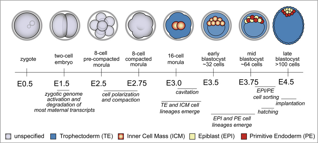
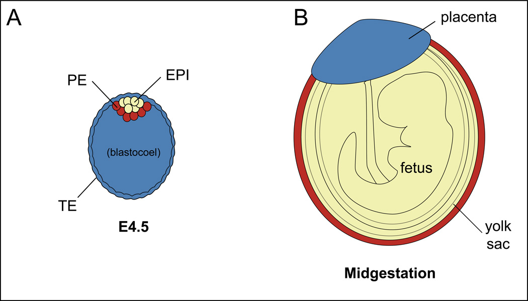
The first cell fate decisions during mammalian development establish tissues essential for healthy pregnancy. The mouse has served as a valuable model for discovering pathways regulating the first cell fate decisions because of the ease with which early embryos can be recovered and the availability of an arsenal of classical and emerging methods for manipulating gene expression. We summarize the major pathways that govern the first cell fate decisions in mouse development. This knowledge serves as a paradigm for exploring how emergent properties of a self-organizing system can dynamically regulate gene expression and cell fate plasticity. Moreover, it brings to light the processes that establish healthy pregnancy and ES cells. We also describe unsolved mysteries and new technologies that could help to overcome experimental challenges in the field.

Keywords: emergent properties; gene networks; pluripotency; preimplantation; reproduction; stem cells.


Figures

Figure 1
Figure 1. Developmental milestones prior to implantation
Cell fates are color-coded according to the legend at the bottom of the figure, and E refers to the number of days after fertilization, which occurs at E0.0. Zygotic genome activation occurs at the two-cell stage, concurrent with degradation of the maternally provided transcriptome. Cell polarization and compaction occur at the late eight-cell stage. TE and ICM cell fates emerge during the eight to sixteen cell transition, and then the blastocoel forms by cavitation, starting between E3.0 and E3.25. During blastocyst expansion, the TE exists as a polarized epithelium, while ICM cells are nonpolar. As the ICM matures, EPI and PE cell fates emerge in the absence of an apparent spatial pattern. Eventually, EPI and PE cells occupy discrete layers within the ICM. Implantation occurs around E4.5, after the embryo hatches from the zona pellucida (grey ring encircling the cleavage stage embryos).
Figure 2
Figure 2. Destinies of the blastocyst lineages
A) At E4.5, the blastocyst contains three cell types, corresponding to three major lineages: one fetal, and two extraembryonic. B) The three major lineages are color-coded in the later mouse embryo, according to their cell type of origin in the blastocyst.
Figure 3
Figure 3. Initiation and maintenance of TE and ICM cell fates
A) Beginning around the 16-cell stage, the HIPPO pathway is active in the ICM, where it represses expression of TE genes, such as Cdx2 and Sox2. The HIPPO pathway is inactive in the TE, enabling the transcriptional regulation of both ICM and TE genes in parallel (WWTR1 omitted for simplicity, please see text for details). B) At later stages, CDX2 reinforces the first cell fate decision by regulating expression of later-acting TE and ICM genes, such as Eomes and Oct4/Nanog, and GATA6 also helps repress Nanog in the TE. Simultaneously, OCT4 helps repress TE genes in some ICM cells. Please see text for relevant citations.
Figure 4
Figure 4. Origins of cellular heterogeneity in the ICM
A) At the onset of blastocyst formation, Fgf4 and Fgfr2 transcripts exhibit heterogeneous distribution within the ICM. Bi) Around the 64-cell stage (E3.75), EPI factors promote expression of FGF4, which is received and transduced by FGFR2/MAPK. FGF4/MAPK signaling increases expression of GATA6, which represses NANOG and promotes expression of PE genes in PE cells. In EPI cells, NANOG represses expression of GATA6. Bii) In both EPI and PE cells, OCT4 is transcriptionally active, but the choice of targets depends on the presence of either SOX2 or SOX17. Please see text for relevant citations.

References

    1. Stephenson M, Kutteh W. Evaluation and management of recurrent early pregnancy loss. Clin Obstet Gynecol. 2007;50:132–145. - PubMed
    1. Molls M, et al. A comparison of the cell kinetics of pre-implantation mouse embryos from two different mouse strains. Cell Tissue Kinet. 1983;16:277–283. - PubMed
    1. Boroviak T, et al. The ability of inner-cell-mass cells to self-renew as embryonic stem cells is acquired following epiblast specification. Nat Cell Biol. 2014;16:516–528. - PMC - PubMed
    1. Parfitt DE, Shen MM. From blastocyst to gastrula: gene regulatory networks of embryonic stem cells and early mouse embryogenesis. Philos Trans R Soc Lond B Biol Sci. 2014;369 - PMC - PubMed
    1. Ralston A, Rossant J. Genetic regulation of stem cell origins in the mouse embryo. Clin Genet. 2005;68:106–112. - PubMed

Publication types

Substances