Review
. 2015 Jun;29(2):357-70.
doi: 10.1016/j.idc.2015.02.008.

Babesiosis

Affiliations
Review

Babesiosis

Edouard G Vannier et al. Infect Dis Clin North Am. 2015 Jun.

Abstract

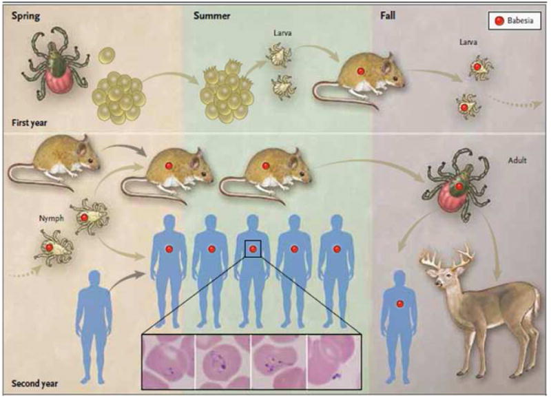
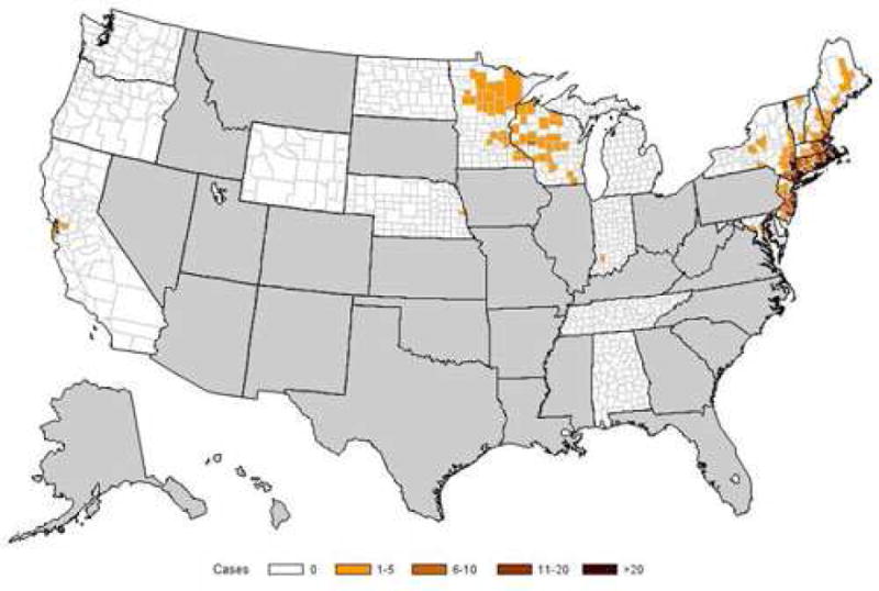
Babesiosis is caused by intraerythrocytic protozoan parasites that are transmitted by ticks, or less commonly through blood transfusion or transplacentally. Human babesiosis was first recognized in a splenectomized patient in Europe but most cases have been reported from the northeastern and upper midwestern United States in people with an intact spleen and no history of immune impairment. Cases are reported in Asia, Africa, Australia, Europe, and South America. Babesiosis shares many clinical features with malaria and can be fatal, particularly in the elderly and the immunocompromised.

Keywords: Apicomplexa; Babesia microti; Babesiosis; Erythrocyte; Protozoan; Tick; Transfusion.


Figures

Figure 1
Figure 1. Transmission of Babesia microti by the Ixodes scapularis tick
Adult female ticks lay eggs in the spring (first year, top left panel). Larvae hatch in the early summer and become infected with B. microti (red circle) as they take a blood meal from infected white-footed mice (Peromyscus leucopus) in late summer. White-footed mice are the primary reservoir host, but other small rodents may carry B. microti. Larvae molt to nymphs the following spring (second year, bottom left panel). When infected nymphs feed on mice or humans in late spring or early summer, these hosts may become infected. Humans are incidental hosts. In the fall, nymphs molt to adults that feed on white-tailed deer (Odocoileus virginianus) but rarely on humans. White-tailed deer do not become infected with B. microti but amplify the tick population by providing a blood meal for adult ticks. The following spring, adult female ticks lay eggs and the cycle is repeated. B. microti are obligate parasites of erythrocytes and typically are visualized on a Giemsa stained thin blood smear (see inset).- The inset panels from left to right show a ring form with a non-staining vacuole surrounded by cytoplasm (in blue) and a small nucleus (in purple), an amoeboid form, a tetrad (also referred to as Maltese Cross), and an extracellular form. From Vannier E, Krause PJ. Human babesiosis. New Engl J Med 2012;366:2397-407 (reference 2).
Figure 2
Figure 2. Geographic distribution of human cases of babesiosis in the United States
Babesiosis became a nationally notifiable condition in January 2011. As of 2012, babesiosis is reportable in 22 states and the District of Columbia. The figure shows the incidence of babesiosis (number of cases per 100,000 persons) by county of residence in 2012. Human babesiosis caused by B. microti has long been reported from the Northeast, particularly from Massachusetts to New Jersey, and recently became endemic in northern New England (Maine and New Hampshire) and in the northern Mid-Atlantic States (from Pennsylvania to Maryland). B. microti also causes disease in the upper Midwest, particularly in Wisconsin and Minnesota. B. duncani has been the etiologic agent along the northwest Pacific Coast. Cases of B. divergens-like infection have been reported from Washington State, Kentucky and Missouri. Adapted from Centers for Disease Control and Prevention (http://www.cdc.gov/parasites/babesiosis/data-statistics.html).
Figure 3
Figure 3. Algorithm for diagnosis of human babesiosis caused by Babesia microti
The laboratory diagnosis of babesiosis should only be initiated for patients who are at risk of infection and for whom there is strong suspicion of infection., Laboratory testing is required for definitive diagnosis of babesiosis. From Vannier E, Krause PJ. Human babesiosis. New Engl J Med 2012;366:2397-407 (reference 2).

References

    1. Skrabalo A, Deanovic A. Piroplasmosis in man: Report on a case. Doc Med Geogr Trop. 1957;9:11–16. - PubMed
    1. Vannier E, Krause PJ. Human babesiosis. New Engl J Med. 2012;366:2397–407. - PubMed
    1. Hunfeld KP, Hildebrandt A, Gray JS. Babesiosis: recent insights into an ancient disease. Int J Parasitol. 2008;38:1219–37. - PubMed
    1. Spielman A, Wilson ML, Levine JF, et al. Ecology of Ixodes dammini borne human babesiosis and Lyme disease. Ann Rev Entomol. 1985;30:439–60. - PubMed
    1. Cornillot E, Hadj-Kaddour K, Dassouli A, et al. Sequencing of the smallest Apicomplexan genome from the human pathogen Babesia microti. Nucleic Acids Res. 2012:1–13. - PMC - PubMed

Publication types

MeSH terms