Review
. 2015 May 22;116(11):1820-34.
doi: 10.1161/CIRCRESAHA.116.305420.

Maintaining ancient organelles: mitochondrial biogenesis and maturation

Affiliations
Review

Maintaining ancient organelles: mitochondrial biogenesis and maturation

Rick B Vega et al. Circ Res. .

Abstract

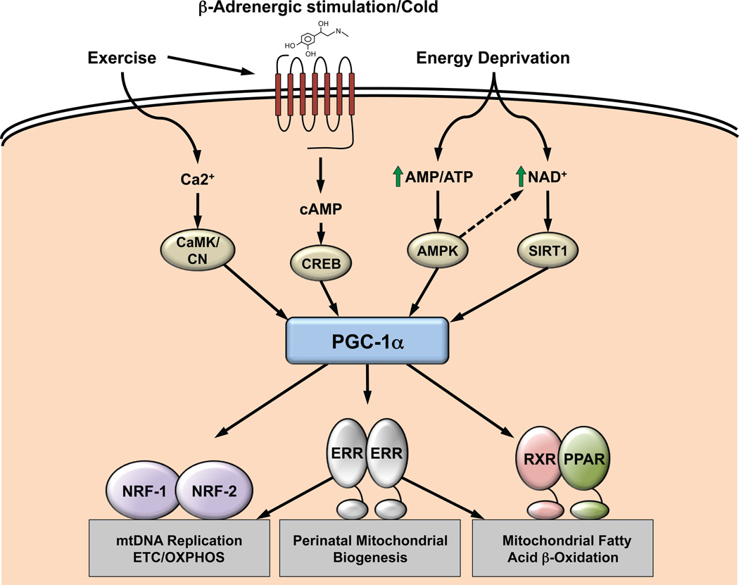
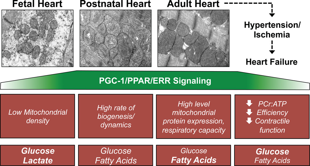
The ultrastructure of the cardiac myocyte is remarkable for the high density of mitochondria tightly packed between sarcomeres. This structural organization is designed to provide energy in the form of ATP to fuel normal pump function of the heart. A complex system comprised of regulatory factors and energy metabolic machinery, encoded by both mitochondrial and nuclear genomes, is required for the coordinate control of cardiac mitochondrial biogenesis, maturation, and high-capacity function. This process involves the action of a transcriptional regulatory network that builds and maintains the mitochondrial genome and drives the expression of the energy transduction machinery. This finely tuned system is responsive to developmental and physiological cues, as well as changes in fuel substrate availability. Deficiency of components critical for mitochondrial energy production frequently manifests as a cardiomyopathic phenotype, underscoring the requirement to maintain high respiration rates in the heart. Although a precise causative role is not clear, there is increasing evidence that perturbations in this regulatory system occur in the hypertrophied and failing heart. This review summarizes current knowledge and highlights recent advances in our understanding of the transcriptional regulatory factors and signaling networks that serve to regulate mitochondrial biogenesis and function in the mammalian heart.

Keywords: mitochondria; mitochondrial turnover; myocytes, cardiac; oxidative phosphorylation; transcription factors.


Figures

Figure 1
Figure 1. The two predominant models of mtDNA replication are shown here
Both models concur the replisome consists of at least a helicase, TWINKLE (orange) and POLG (yellow). Leading strand synthesis begins at OH and lagging strand synthesis at OL (red arrow). A) Strand-displacement model (SDM) proposes SSB proteins (green spheres) bind the displaced H-strand during leading strand replication. B) Alternatively, the ribonucleotide incorporation throughout the lagging strand (RITOLS) model proposes portions of transcribed mtDNA bind the H-strand (green dotted line).
Figure 2
Figure 2. POLMRT Plays a Critical Role in Mitochondrial Transcription and Replication
A) The transcription pre-initiation begins with mitochondrial transcription factor A (TFAM) binding and recruiting POLMRT. TFAM enables POLMRT interaction with upstream promoter (P) by bending the DNA around POLMRT. B) Transcription initiation occurs when TF2BM binds POLMRT and facilitates promoter melting forming the characteristic D-loop region. POLMRT synthesizes an RNA primer (green dotted line) until reaching CSBII where the transcription/replication switch occurs. C) In the presence of TEFM the G-quadruplex that stalls POLMRT is disrupted allowing POLMRT to continue adding nucleotides and completing transcription (top). In the absence of TEFM, POLMRT disassociates from mtDNA, transcription is terminated at CSBII and the oligonucleotide strand is used as a primer for DNA replication (bottom). Replication then proceeds following the recruitment of the replisome apparatus. TFAM, mitochondrial transcription factor A; POLMRT, mitochondrial RNA polymerase; TFB2M, mitochondrial transcription factor B2; CSBII, conserved sequence block II; TEFM, mitochondrial transcription elongation factor.
Figure 3
Figure 3. PGC-1α mediates physiologic control of mitochondrial biogenesis and function
The transcriptional coactivator PGC-1α interacts directly with multiple transcription factors to integrate upstream signaling events with mitochondrial biogenesis and functional capacity. The downstream transcription factors control virtually every aspect of mitochondrial function and energy production including biogenesis, dynamics, and maintenance of protein levels. The control of PGC-1α expression and activity is dynamic, responding to multiple intracellular second messengers and signaling molecules transmitting inputs from various physiologic and metabolic stimuli (top). CREB, cAMP-response element binding protein; AMPK, AMP-activated protein kinase; SIRT1, sirtuin 1; CaMK, calmodulin-dependent kinase; CN, calcineurin; NAD+, nicotinamide adenine dinucleotide; PGC-1α, PPARγ coactivator 1; PPAR, peroxisome proliferator-activated receptor; NRF, nuclear respiratory factor; ERR, estrogen-related receptor; ETC/OXPHOS, electron transport chain/oxidative phosphorylation.
Figure 4
Figure 4. Dynamic changes in cardiac mitochondrial number, structure, and function during developmental stages and in the failing heart
Electron micrographs reveal a developmentally regulated increase in cardiac myocyte mitochondrial number and organization during the transition from the fetal period to the adult. The surge of mitochondrial biogenesis at birth and subsequent maturation in the postnatal heart is driven by induction of PGC-1. In the adult heart, PGC-1 maintains high level, coordinated expression of nuclear and mitochondrial genes encoding mitochondrial machinery. Mitochondrial function and energy production is compromised in the failing heart concomitant with decreased ERR/PPAR/PGC-1 signaling. PPAR, peroxisome proliferator-activated receptor; ERR, estrogen-related receptor; PGC-1, PPARγ coactivator 1; PCr, phosphocreatine.

References

    1. Miller FJ, Rosenfeldt FL, Zhang C, Linnane AW, Nagley P. Precise determination of mitochondrial DNA copy number in human skeletal and cardiac muscle by a PCR-based assay: lack of change of copy number with age. Nucleic Acids Res. 2003;31:e61. - PMC - PubMed
    1. Pagliarini DJ, Calvo SE, Chang B, et al. A mitochondrial protein compendium elucidates complex I disease biology. Cell. 2008;134:112–123. - PMC - PubMed
    1. Huang X, Sun L, Ji S, Zhao T, Zhang W, Xu J, Zhang J, Wang Y, Wang X, Franzini-Armstrong C, Zheng M, Cheng H. Kissing and nanotunneling mediate intermitochondrial communication in the heart. Proc Natl Acad Sci U S A. 2013;110:2846–2851. - PMC - PubMed
    1. Picard M, McManus MJ, Csordas G, Varnai P, Dorn GW, 2nd, Williams D, Hajnoczky G, Wallace DC. Trans-mitochondrial coordination of cristae at regulated membrane junctions. Nat Commun. 2015;6:6259. - PMC - PubMed
    1. Robberson DL, Kasamatsu H, Vinograd J. Replication of mitochondrial DNA. Circular replicative intermediates in mouse L cells. Proc Natl Acad Sci U S A. 1972;69:737–741. - PMC - PubMed

Publication types

Substances