Review
. 2015 May 22;116(11):1863-82.
doi: 10.1161/CIRCRESAHA.116.305372.

Proteotoxicity and cardiac dysfunction

Affiliations
Review

Proteotoxicity and cardiac dysfunction

Patrick M McLendon et al. Circ Res. .

Abstract

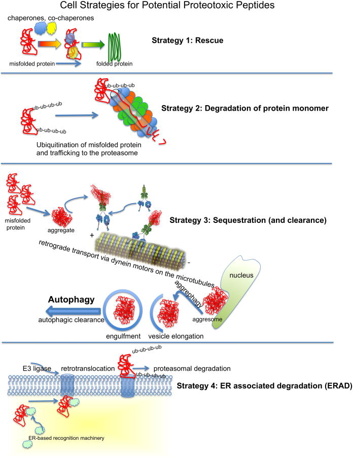
Baseline physiological function of the mammalian heart is under the constant threat of environmental or intrinsic pathological insults. Cardiomyocyte proteins are thus subject to unremitting pressure to function optimally, and this depends on them assuming and maintaining proper conformation. This review explores the multiple defenses a cell may use for its proteins to assume and maintain correct protein folding and conformation. There are multiple quality control mechanisms to ensure that nascent polypeptides are properly folded and mature proteins maintain their functional conformation. When proteins do misfold, either in the face of normal or pathological stimuli or because of intrinsic mutations or post-translational modifications, they must either be refolded correctly or recycled. In the absence of these corrective processes, they may become toxic to the cell. Herein, we explore some of the underlying mechanisms that lead to proteotoxicity. The continued presence and chronic accumulation of misfolded or unfolded proteins can be disastrous in cardiomyocytes because these misfolded proteins can lead to aggregation or the formation of soluble peptides that are proteotoxic. This in turn leads to compromised protein quality control and precipitating a downward spiral of the cell's ability to maintain protein homeostasis. Some underlying mechanisms are discussed and the therapeutic potential of interfering with proteotoxicity in the heart is explored.

Keywords: autophagy; proteasome; protein.


Figures

Figure 1
Figure 1
Schematic diagram of selected protection strategies or proteotoxic pathways in the cardiomyocyte. Figure 1. Schematic diagram of selected protection strategies in the cardiomyocyte for dealing with misfolded or potentially proteotoxic peptides. Strategy 1) [Rescue], the cell's chaperones, which can be constitutively expressed or inducible, act alone or in concert to maintain or restore the peptide's functional conformation. Strategy 2) [Degradation], the misfolded protein is recognized by the Protein Quality Control machinery, ubiquitinated and trafficked to the proteasome for degradation and amino acid recycling. Strategy 3) [Sequestration], the misfolded protein forms larger aggregates, which are unable to be processed by the proteasome. These aggregates can be attached to dynein motors and transported on microtubules to a perinuclear location where the potentially proteotoxic peptides are sequestered. These aggregates can be cleared from the cytoplasm by autophagy (see text for details).,, Strategy 4) [ERAD] The endoplasmic reticulum associated degradation pathway is quite complex and a simplified schematic diagram simply outlines the process. Mutated or terminally misfolded proteins are recognzied by ER assocaited proteins that may have either enzymatic or chaperone activities. The protein is then sheparhaerded to the retrotranslocation machinery whoese exact identity(ies) remain obscure. Membrane associated E3 ligases and associated proteins are able to mediate ubiquitination of the protein, which may partially drive its retrotranslocation. There are multiple checkpoints and components underlying these processes as well (see text for details). The ubiquitinated proteins are then targeted for proteasomal degradation.
Figure 2
Figure 2
Selective cargo recognition and degradation by autophagy. Step 1, Ubiquitin binding is necessary for most modes of selective autophagy. Polyubiquitin conjugation onto a substrate protein is accomplished via an E3 ubiquitin ligase. Many of these proteins are ubiquitinated via a K63 linkage, which signals lysosomal degradation rather than the K48 linkage that is typical for proteasomal targeting. Step 2, Ubiquitinated proteins can be recognized by cargo receptors, such as p62, NBR1 and/or HDAC6 for transport towards the lysosome or aggresome. p62 ( formula image
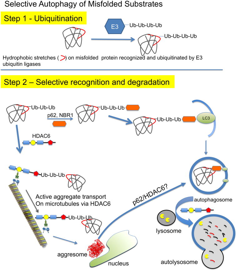
) binds ubiquitinated proteins for interaction with LC3 in a growing autophagosome, thus selectively targeting the protein for autophagic degradation after fusion with lysosomes. In addition, HDAC6 can bind ubiquitinated proteins through its ubiquitin binding domain ( formula image) and transport them in a retrograde fashion along microtubules by binding to the motor protein dynein through the dynein motor binding domain ( formula image). These can be single misfolded species or soluble proto-aggregates. This event can happen many times, causing the proteins to coalesce and form aggresomes in perinuclear region of the cell. Aggresomal proteins can also be degraded by autophagy, and these cargo transport proteins may be involved in this process as well, particularly HDAC6 with known roles in mediating the autophagosome-lysosome fusion event.
Figure 3
Figure 3
Anatomy of aggregates. A, Shown is a typical, mature aggregate in a CryABR120G cardiomyocyte derived from a 4 month old, symptomatic heart. Note the fenestrated organization and the mitochondria trapped within the aggregate. B, Immunogold staining (dark grains) showing the strong presence of CryAB within both the large, well defined aggregates and the smaller, more amorphous masses, some of which are mitochondria (mit). C, D, High magnification, immunogold staining using cardiomyocytes derived from 6 week old CryABR120G hearts showing the internalization of CryAB, but also (D) the presence of desmin. mit; mitochondria. Photomicrographs courtesy of H. Osinska. Mice, fixation techniques and details of the transmission electron microscopy were carried out as described.
Figure 4
Figure 4
Proteotoxic peptides. Shown is the formation of what are thought to be the toxic entities in a wide variety of proteotoxic diseases; small, soluble pre-amyloid oligomers. These are metastable entities in slow or rapid equilibriums with one another and, at least for some entities, the dimers and trimers are the most toxic form. Enhanced autophagy is correlated with reduction in toxic pre-amyloid levels but the mechanism of clearance remains obscure (see text for details).
Figure 5
Figure 5
Autophagy PCR array in voluntary exercised CryABR120GxAtg7xtTA hearts. Graph representing direct group wise comparison of fold change in mRNA levels in male voluntary exercise CryABR120GxAtg7xtTA and control non-exercised CryABR120GxAtg7xtTA hearts at 4.5 months (fold change versus non-exercised CryABR120GxAtg7xtTA control, n = 3 per group). All values are reported as mean ± S.E.M. P<0.05 and P<0.01 by Student's t test. Mice and experimental treatments were carried out as described.previously.
Figure 6
Figure 6
High-throughput assay to uncover novel effectors of cardiac protein aggregation. A, Screening Principle. A cell model of cardiac proteotoxicity was developed in primary cardiomyocytes. These cells were subjected to systematic, genome-wide knockdown by infection with lentiviruses expressing shRNAs capable of being processed such that selective degradation of their cognate mRNAs took place. The goal of the screen is to find genes that, when knocked down, lead to a reduction of aggregates. B, An example of a candidate gene or “hit” in the screen. The top panels show the images taken from one well, in which 16 images are acquired and aggregate content quantified. The lower panel depicts one image from a well. In the well treated with siRNA, a drastic reduction in aggregate content is observed compared to controls. This figure demonstrates the very high signal to noise ratio observed upon expression of CryABR120G, which allows one to robustly quantitate changes in aggregate content. Scale bars: top panel, 1000 μm; bottom panel, 200 μm.

References

    1. Galluzzi L, Bravo-San Pedro JM, Vitale I, et al. Essential versus accessory aspects of cell death: Recommendations of the nccd 2015. Cell Death Differ. 2015;22:58–73. - PMC - PubMed
    1. Kroemer G, Galluzzi L, Vandenabeele P, et al. Classification of cell death: Recommendations of the nomenclature committee on cell death 2009. Cell Death Differ. 2009;16:3–11. - PMC - PubMed
    1. Hightower LE. Heat shock, stress proteins, chaperones, and proteotoxicity. Cell. 1991;66:191–197. - PubMed
    1. Douglas PM, Cyr DM. Interplay between protein homeostasis networks in protein aggregation and proteotoxicity. Biopolymers. 2010;93:229–236. - PMC - PubMed
    1. Christie NT, Lee AL, Fay HG, Gray AA, Kikis EA. Novel polyglutamine model uncouples proteotoxicity from aging. PLoS One. 2014;9:e96835. - PMC - PubMed

Publication types

LinkOut - more resources