. 2015 May 22;348(6237):880-6.
doi: 10.1126/science.aaa6806.

Tumor evolution. High burden and pervasive positive selection of somatic mutations in normal human skin

Affiliations

Tumor evolution. High burden and pervasive positive selection of somatic mutations in normal human skin

Iñigo Martincorena et al. Science. .

Abstract

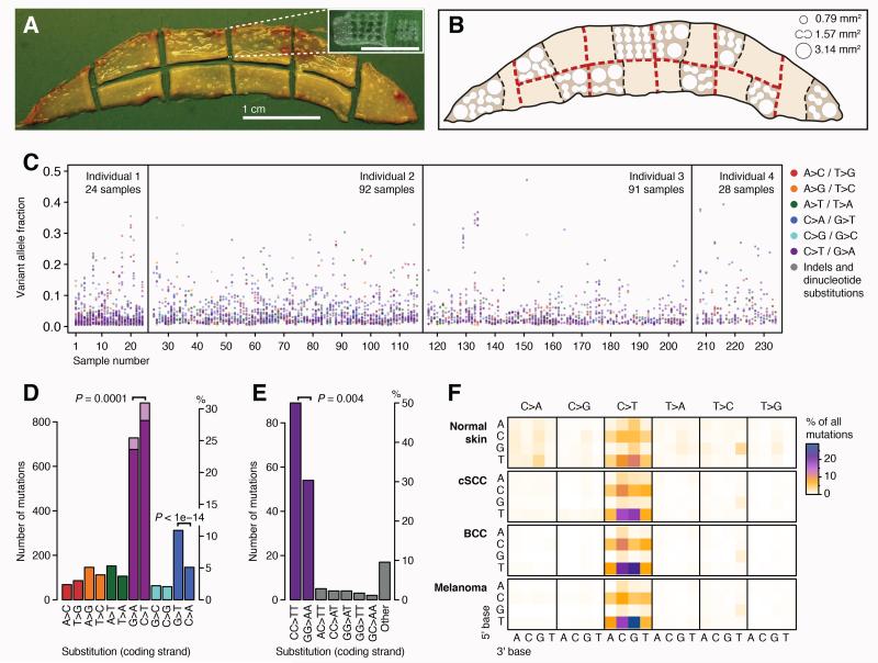
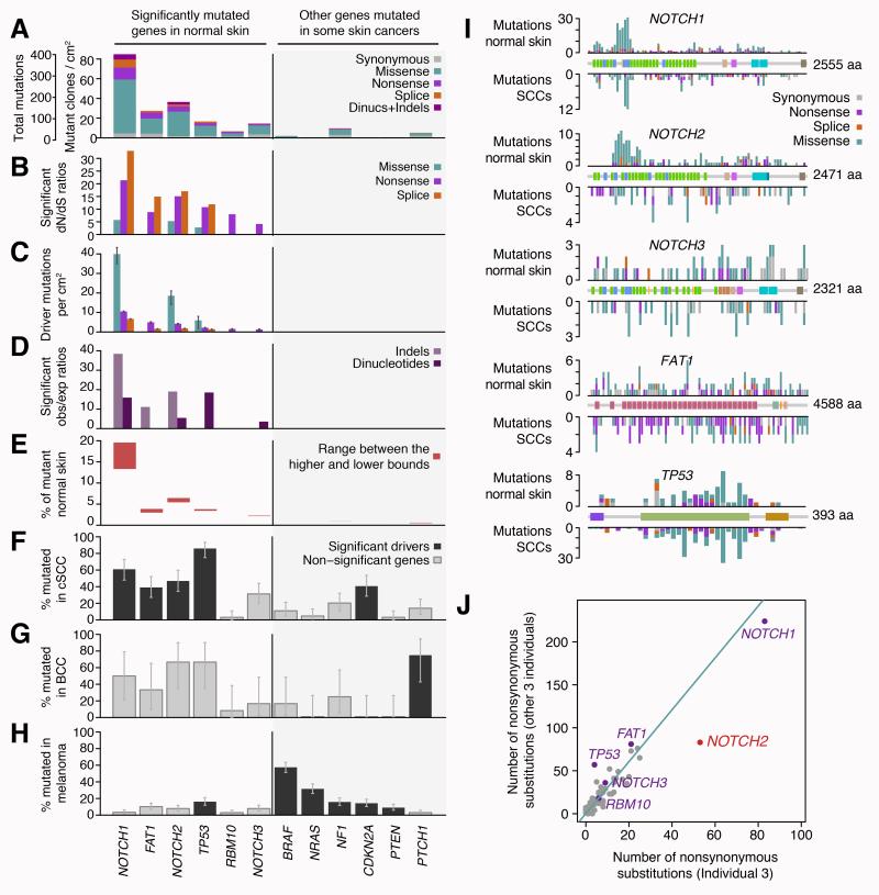
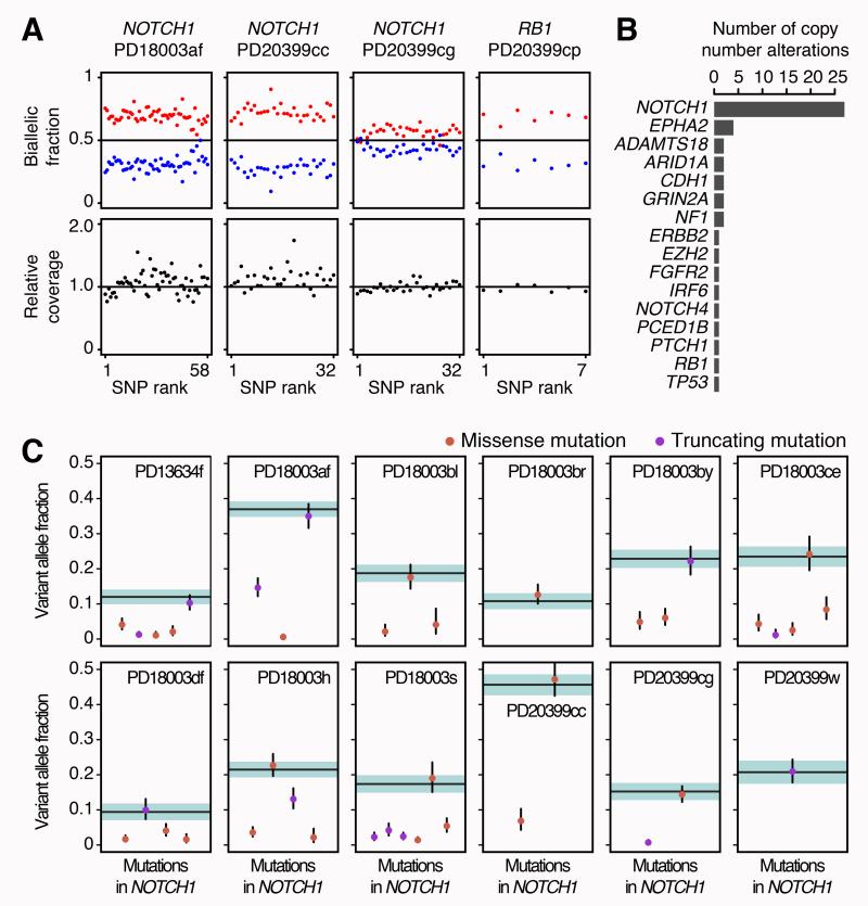
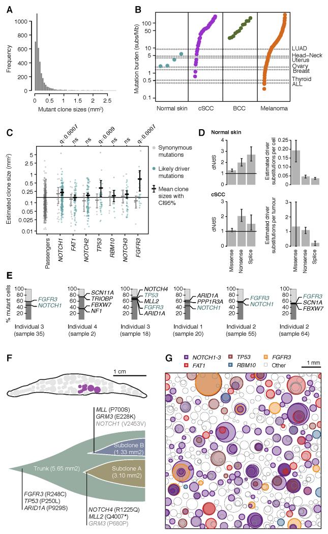
How somatic mutations accumulate in normal cells is central to understanding cancer development but is poorly understood. We performed ultradeep sequencing of 74 cancer genes in small (0.8 to 4.7 square millimeters) biopsies of normal skin. Across 234 biopsies of sun-exposed eyelid epidermis from four individuals, the burden of somatic mutations averaged two to six mutations per megabase per cell, similar to that seen in many cancers, and exhibited characteristic signatures of exposure to ultraviolet light. Remarkably, multiple cancer genes are under strong positive selection even in physiologically normal skin, including most of the key drivers of cutaneous squamous cell carcinomas. Positively selected mutations were found in 18 to 32% of normal skin cells at a density of ~140 driver mutations per square centimeter. We observed variability in the driver landscape among individuals and variability in the sizes of clonal expansions across genes. Thus, aged sun-exposed skin is a patchwork of thousands of evolving clones with over a quarter of cells carrying cancer-causing mutations while maintaining the physiological functions of epidermis.


Figures

Fig. 1
Fig. 1. Burden and spectrum of mutations in normal human skin
(A) Excised human eyelid viewed from the dermal surface. The inset shows a sample region of epidermis after the dermis has been removed and biopsies taken. (B) Locational map of harvested areas from an eyelid showing locations of 0.79mm2, 1.57mm2 and 3.14mm2 biopsies. (C) Distribution of the variant allele fraction (i.e. the fraction of sequencing reads reporting the mutation of all reads across the locus) for the 3,760 mutations found across the 234 samples from 4 individuals, colored by mutation type. (D, E) Total counts in the coding (untranscribed) versus the non-coding (transcribed) strand for single base substitutions (D) and dinucleotides (E). The counts of C>T (G>A) mutations in a dipyrimidine context are shown in dark purple. P-values reflect the transcription strand asymmetry (Exact Poisson test). (F) Heat map of the relative rates of each mutation type depending on the nucleotides upstream and downstream of the mutated base. Rates are normalized for sequence composition of the targeted genes.
Fig. 2
Fig. 2. Pervasive positive selection of oncogenic mutations in normal skin
(A-E) Patterns of selection in six genes recurrently mutated in normal skin and in six other genes frequently implicated in skin cancers. (A) Number of mutations per gene classified by their functional impact. (B) dN/dS ratios for genes under significant positive selection (only statistically significant ratios are shown). (C) Estimated number of driver mutations per cm2 of normal skin. (D) Enrichment of indels and dinucleotides in driver genes (bars show significant observed/expected ratios only). (E) Estimated percentage of cells in normal skin carrying mutations in each gene. Lower bound estimates were obtained assuming the possibility of up to two driver mutations per cell, while higher bound estimates are obtained by allowing only one driver mutation per gene per cell. (F-H) Percentage of cSCC, BCC and melanoma tumors that carry a non-synonymous point mutations in each gene. Genes found to be recurrently mutated in each cancer type are shown in black (supplementary results S2.2). (I) Distribution of mutations across five driver genes in normal skin (above the gene diagrams) and in SCCs (below), including 67 cutaneous SCCs and 319 TCGA head and neck cancer exomes. The gene diagrams show the location of encoded protein domains. (J) Differential selection in NOTCH2 across individuals (supplementary methods S1.5).
Fig. 3
Fig. 3. Frequent copy number aberrations and biallelic loss of NOTCH1 in normal skin
(A) Example of four skin samples with subclonal copy number aberrations in NOTCH1 and RB1. Every point represents a heterozygous SNP within the affected gene and aberrations manifest as allelic imbalances, with a higher fraction of reads (biallelic fraction) supporting one of the alleles of the gene (in red). The extent of the deviation from 0.5 depends on the number of gene copies gained or lost and on the proportion of the biopsy occupied by the subclone (supplementary methods S1.6). (B) Number of copy number aberrations detected per gene. (C) In NOTCH1, a substitution is often found in the same fraction of cells as a deletion of the other allele (dot colocalizing with a horizontal band), showing that the loss of both copies of NOTCH1 is frequent in normal skin cells. Horizontal lines represent the expected variant allele fraction for a mutation inactivating the only remaining allele of a gene in the same clone, with colored shadows representing 95% confidence intervals. Orange and purple dots represent the allele fraction of missense and nonsense mutations in the biopsy, with 95% confidence intervals (supplementary methods S1.6.1, Fig. S5).
Fig. 4
Fig. 4. Mutant clone sizes and clonal dynamics in normal skin
(A) Distribution of clone sizes of all mutations. (B) Mutation burden per megabase in the normal skin of four individuals and in a range of human cancers (supplementary methods S1.8). (C) Clone sizes of likely driver and passenger mutations in normal skin. Driver mutations are defined as those mutation types found to be under significant positive selection in each gene (Fig. 2). Confidence intervals and FDR-corrected q-values were obtained using 10,000 random permutations of the gene labels assigned to each mutation. (D) Global dN/dS estimates across all 74 genes analyzed in the study in normal skin and cSCC. This allows us to estimate the number of driver mutations per normal cell or per tumor as the number of mutations fixed by positive selection (supplementary methods S1.4.2). (E) Identification of mutations co-occurring in the same sub-clone using the pigeonhole principle (32). (F) Subclonal structure of a large clone found to overlap with six biopsies (shown in purple in the eyelid locational map). (G) Schematic representation of the mutant clones in an average 1 cm2 of normal eyelid skin. To generate the figure, a number of biopsies were randomly selected to amount to 1cm2 of sequenced skin and all clones observed in these biopsies were represented as circles randomly distributed in space. The density, size and the simulated nesting of clones are all based on the sequencing data obtained in this study.

Comment in

  • Cancer. Preprocancer.
    Brash DE. Brash DE. Science. 2015 May 22;348(6237):867-8. doi: 10.1126/science.aac4435. Science. 2015. PMID: 25999495 No abstract available.

References

    1. Vogelstein B, et al. Cancer genome landscapes. Science. 2013 Mar 29;339:1546. - PMC - PubMed
    1. Garraway LA, Lander ES. Lessons from the cancer genome. Cell. 2013 Mar 28;153:17. - PubMed
    1. Tomasetti C, Vogelstein B, Parmigiani G. Half or more of the somatic mutations in cancers of self-renewing tissues originate prior to tumor initiation. Proceedings of the National Academy of Sciences of the United States of America. 2013 Feb 5;110:1999. - PMC - PubMed
    1. Nowell PC. The clonal evolution of tumor cell populations. Science. 1976 Oct 1;194:23. - PubMed
    1. Jackson AL, Loeb LA. The mutation rate and cancer. Genetics. 1998 Apr;148:1483. - PMC - PubMed

Publication types

MeSH terms