Review
. 2015 May 22;348(6237):aaa6566.
doi: 10.1126/science.aaa6566. Epub 2015 May 21.

Innate lymphoid cells. Innate lymphoid cells: a new paradigm in immunology

Affiliations
Review

Innate lymphoid cells. Innate lymphoid cells: a new paradigm in immunology

Gérard Eberl et al. Science. .

Abstract

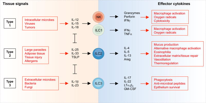
Innate lymphoid cells (ILCs) are a growing family of immune cells that mirror the phenotypes and functions of T cells. However, in contrast to T cells, ILCs do not express acquired antigen receptors or undergo clonal selection and expansion when stimulated. Instead, ILCs react promptly to signals from infected or injured tissues and produce an array of secreted proteins termed cytokines that direct the developing immune response into one that is adapted to the original insult. The complex cross-talk between microenvironment, ILCs, and adaptive immunity remains to be fully deciphered. Only by understanding these complex regulatory networks can the power of ILCs be controlled or unleashed in order to regulate or enhance immune responses in disease prevention and therapy.


Figures

Figure 1
Figure 1. The development of ILCs
The development of ILCs from common lymphoid progenitors (CLPs) requires Id2-mediated suppression of alternative lymphoid cell fates that generate B and T cells. Factors present in the microenvironment, such as Notch ligands, bone morphogenic proteins (BMPs) and cytokines, as well as the circadian rhythm, control expression of Nfil3, Gata3 and Id2, which determine the progression towards the ILC fate. Distinct precursors give rise to NK cells and ILCs (which, unlike NK cells, are non-cytotoxic), while the transcription factor PLZF further divides the progeny of ChILPs into the PLZF-dependent ILC1s, ILC2s and ILC3s, and PLZF-independent LTi cells (although LTi cells tend to be grouped as ILC3s) required for the development of lymph nodes, Peyer’s patches and ILFs. The maturation of ILC precursors into mature ILCs may occur outside of primary lymphoid tissues, in ways similar to the maturation of naïve T helper cells into Th1, Th2, Th17 and Treg cells, and in response to a variety of signals produced by the tissue microenvironment.
Figure 2
Figure 2. Activation and functions of ILCs
The tissue signals that expand and activate ILC1s, ILC2s and ILC3s, and the effector functions of ILCs, mirror the activation and functions of T cells. In this figure, NK cells, ILC1s, ILC2s and ILC3s could be replaced by CD8+ T cells, Th1, Th2 and Th17 cells, respectively. However, while ILCs are activated promptly by tissue signals and therefore act upstream in the immune response, T cells are first selected and expanded on the basis of T cell receptor specificity, a process that typically requires several days.
Figure 3
Figure 3. Regulation of adaptive immunity by ILCs
ILCs regulate T cells both directly through antigen presentation on MHC II, and indirectly through the regulation of DCs. The crosstalk between ILCs, DCs and T cells establishes a complex regulatory network, involving positive and negative feedbacks, the dynamics of which remain to be elucidated. The mechanisms by which ILCs repress CD4+ T helper (Th) cell activation remain unclear, but may involve the lack of costimulatory molecules in the context of steady-state (84). It also remains unclear how DCs negatively regulate the activity of ILCs (57). Red lines depict feedback loops, and A, B and C list the type 1, type 2 or type 3 cytokines involved in a specific crosstalk. ILC3s also activate B cells in the intestine through lymphotoxin-mediated recruitment of T helper cells and activation of dendritic cells (91), as well as marginal zone B cells in the spleen (132).
Figure 4
Figure 4. ILCs in pathology
Pathogens, allergens, chemicals, diet, metabolic states and genetic factors can induce type 1, type 2 or type 3 inflammatory conditions that lead to pathology involving ILCs. Listed are examples of pathologies shown to involve ILCs, even though in most cases the causative role of ILCs, or their requirement in the pathology, remains to be established. Strong intestinal inflammatory pathology induced during IBD or by Salmonella enterica generates ILCs that produce both type 1 (IFNγ) and type 3 (IL-17) effector cytokines.

References

    1. Kiessling R, et al. Killer cells: a functional comparison between natural, immune T-cell and antibody-dependent in vitro systems. J Exp Med. 1976;143:772. - PMC - PubMed
    1. Mebius RE, Rennert P, Weissman IL. Developing lymph nodes collect CD4+CD3−LTβ+ cells that can differentiate to APC, NK cells, and follicular cells but not T or B cells. Immunity. 1997;7:493. - PubMed
    1. Adachi S, Yoshida H, Kataoka H, Nishikawa S. Three distinctive steps in Peyer’s patch formation of murine embryo. Int Immunol. 1997;9:507. - PubMed
    1. Cella M, et al. A human natural killer cell subset provides an innate source of IL-22 for mucosal immunity. Nature. 2009;457:722. - PMC - PubMed
    1. Satoh-Takayama N, et al. Microbial flora drives interleukin 22 production in intestinal NKp46+ cells that provide innate mucosal immune defense. Immunity. 2008;29:958. - PubMed

Publication types