Review
. 2015 Jun;64(6):1872-85.
doi: 10.2337/db14-1843.

Human β-cell proliferation and intracellular signaling: part 3

Affiliations
Review

Human β-cell proliferation and intracellular signaling: part 3

Andrew F Stewart et al. Diabetes. 2015 Jun.

Abstract

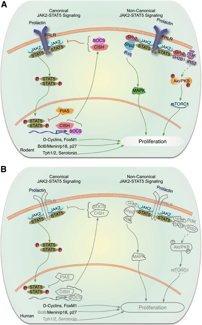
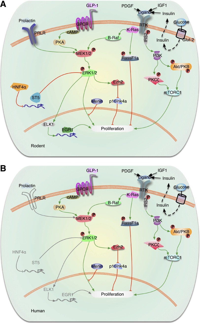
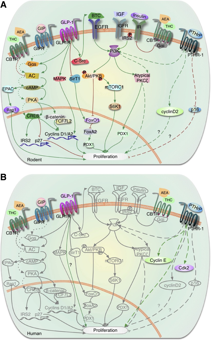
This is the third in a series of Perspectives on intracellular signaling pathways coupled to proliferation in pancreatic β-cells. We contrast the large knowledge base in rodent β-cells with the more limited human database. With the increasing incidence of type 1 diabetes and the recognition that type 2 diabetes is also due in part to a deficiency of functioning β-cells, there is great urgency to identify therapeutic approaches to expand human β-cell numbers. Therapeutic approaches might include stem cell differentiation, transdifferentiation, or expansion of cadaver islets or residual endogenous β-cells. In these Perspectives, we focus on β-cell proliferation. Past Perspectives reviewed fundamental cell cycle regulation and its upstream regulation by insulin/IGF signaling via phosphatidylinositol-3 kinase/mammalian target of rapamycin signaling, glucose, glycogen synthase kinase-3 and liver kinase B1, protein kinase Cζ, calcium-calcineurin-nuclear factor of activated T cells, epidermal growth factor/platelet-derived growth factor family members, Wnt/β-catenin, leptin, and estrogen and progesterone. Here, we emphasize Janus kinase/signal transducers and activators of transcription, Ras/Raf/extracellular signal-related kinase, cadherins and integrins, G-protein-coupled receptors, and transforming growth factor β signaling. We hope these three Perspectives will serve to introduce these pathways to new researchers and will encourage additional investigators to focus on understanding how to harness key intracellular signaling pathways for therapeutic human β-cell regeneration for diabetes.


Figures

Figure 1
Figure 1
Cytokine and hormone signaling through JAK-STAT pathways. In the canonical JAK-STAT signaling cascade, there are four JAKs, 1–3 and TYK2, seven STATs, 1, 2, 3, 4, 5a, 5b, and 6, as well as multiple inhibitors or SOCS1–7, CISH, as well as PIAS. In this complex multimember pathway, cell-type specificity is dictated by which family members are present, and which ones are preferred to others. A: Rodent β-cells. For example, as illustrated for prolactin in rodent β-cells on the left side of the figure showing canonical JAK-STAT signaling, prolactin or placental lactogens bind the dimeric PRLR. PRLR preferentially couples to JAK2, which phosphorylates/activates STAT5a/b. Phosphorylated STAT5a/b dimers then transit to the nucleus and bind to promoters of relevant genes such as cyclin D2 to drive proliferation. Rodent β-cells also contain the dimeric GHR and EPO receptor, which presumably acts via a similar mechanism(s). On the right half of the figure, examples of noncanonical signaling via the PRLR or GHR are illustrated. The major point here is that in addition to activating canonical pathways, cytokines and hormones can also engage MAPK and PI3K signaling pathways discussed in more detail in other figures. B: Human β-cells. Much less is known regarding human β-cell PRL and EPO signaling. PRL and PL fail to activate adult human β-cell proliferation, for reasons that remain unclear. JAK2 and STATs 5a and b are present, as is menin. Several SOCS members, CISH, and PIAS1 are present in human β-cells. Other components of the system present in rodent β-cells, including inhibitors of JAK-STAT signaling, such as SOCS, CISH, and PIAS1, have not been studied. Gray lines are molecules and pathways that are known to exist in rodents but are unknown in human β-cells.
Figure 2
Figure 2
Signaling by Ras/Raf/ERK in the regulation of β-cell proliferation. A: Rodent β-cells. Activation of tyrosine kinase receptors (RTKs) (IGF-1 and paracrine effects of insulin) induces β-cell proliferation by activation of the PI3K/Akt (Akt-protein kinase B)/mTOR and PKCζ and ERK1/2 (also called MAPK) signaling pathways. Recent studies have shown that K-Ras is a major regulator of β-cell proliferation. K-Ras activation regulates β-cell proliferation by modulating two major cascades, B-Raf/MEK/ERK and Rassf1a. Activation of K-Ras signaling activates both the ERK1/2 and the Rassf1a pathways, resulting in suppression of β-cell proliferation when menin is present. This indicates that menin ultimately determines the K-Ras effects on β-cell proliferation downstream of MAPK/ERK signaling. In the case of PDGF, PDGF receptor-α induces ERK1/2 phosphorylation, leading to upregulation of the polycomb group protein Ezh2 and repression of p16INK4a resulting in β-cell expansion. ERK activation also can regulate gene expression by induction of downstream transcription factors, such as ELK1 and Egr1, which in turn regulate expression of proproliferative genes in the nucleus. Other transcription factors, such as HNF-4α, indirectly regulate ERK signaling by downregulation of ST5 under some conditions. Ligand activation of the Gαs subunit of the GLP-1R stimulates adenyl cyclase (AC), which leads to an increase in cAMP and activation of PKA and MEK1/2. B: Human β-cells. Most of the components of the rodent β-cell system are also present in the human β-cell, as illustrated. Exceptions are that PDGF receptor-α is apparently absent in adult human β-cells, and PRLR has not been demonstrated in human β-cells. Gray lines are molecules and pathways that are known to exist in rodents but are unknown in human β-cells.
Figure 3
Figure 3
Cadherin and integrin signaling pathways. A: Rodent β-cells. Activation of β-integrin receptors on β-cells leads to FAK-mediated phosphorylation of Akt/PKB that in turn blocks GSK3β to release the inhibition on β-catenin. The free β-catenin forms a complex with Lef/transcription factor 7–like 2 (Tcf7L2) to promote modulation of cell cycle genes and β-cell proliferation. It is uncertain whether integrins also act via MEK to promote ERK phosphorylation that might in turn activate the β-catenin pathway and/or directly regulate cell cycle genes. E-cadherins are proteins that have been shown to be present in intercellular complexes in islets. Activation of E-cadherins has been linked to the β- and α-catenin complex and promotion of proliferation via the β-catenin modulation of cell cycle genes. B: Human β-cells. Integrins, E-cadherins, and components of the signaling pathways detected in rodents have also been reported to be present in human β-cells. Integrins and E-cadherins have not been shown to be able to modulate proliferation in human β-cells. Gray lines are pathways that are known to exist in rodents but are unknown in human β-cells.
Figure 4
Figure 4
GPCR (or GPR) signaling in the regulation of β-cell proliferation. A: Rodent β-cells. The incretin hormones, GLP-1 and GIP, bind to their respective GPCRs, GLP-1R and GIPR, releasing the “active” Gαs subunit, which stimulates adenylate cyclase (AC) and cAMP synthesis. cAMP signals though either PKA or EPAC pathways. PKA phosphorylates CREB, which upregulates transcription of key proliferative β-cell genes (e.g., cyclin D1, cyclin A2, Pdx1, Irs2) and downregulates transcription of the cell cycle inhibitor p27. IRS2 supports autocrine/paracrine stimulation of IGF/IGF receptor (IGFR)/PI3K-Akt (Akt-PKB) signaling. Akt activation enhances proliferation through a dual effect: activation of mTORC1 and its target p70 ribosomal S6K1 and phosphorylation and inactivation of FOXO1, which allows FOXA activation and PDX1 expression. GLP-1 also inactivates FOXO1 by inhibiting SirT1-mediated deacetylation of FOXO1. Through a variety of cross-talk mechanisms, GLP-1 activates multiple other signaling pathways: PI3K/atypical PKCζ, transactivation of EGFR through c-Src tyrosine kinase activation of its ligand betacellulin (BTC), ERK1/2, and Wnt signaling pathway molecules β-catenin and transcription factor 7–like 2 (TCF7L2). Another GPCR member, PTHR-1, is activated by PTHrP, leading to increased β-cell proliferation through PI3K-atypical PKCζ phosphorylation. Overexpression of PTHrP in β-cells induces cyclin D2 expression and reduces levels of the cell cycle inhibitor p16. Activation of the cannabinoid receptor, CB1R, by its ligands anandamide (AEA) and Δ-9-tetrahydrocannabinol (THC), reduces β-cell proliferation via inhibition of the insulin/IGF-1/IRS2/AKT signaling pathway through the heterotrimeric G-protein, Gαi. The potential effect of CB1R on proliferation by modulating AC is not fully understood. Green arrows indicate activation of pathways and red lines denote inhibition of pathways. Signaling pathways are denoted by solid lines downstream of GLP-1R, dashed lines downstream of PTHR-1, and dotted lines downstream of GIPR. B: Human β-cells. The GPCRs, GLP-1R, GIPR, PTHR-1, and CB1R are all present on human β-cells. Whether GLP-1 enhances human β-cell proliferation is unclear, as one report reveals a modest increase in proliferation (85) and another indicates no increase in replication with GLP-1 analogs (36). PTHrP induces human β-cell proliferation in vitro and increases expression of the cell cycle activators cyclin E and cdk2 in human islets. In gray are molecules and pathways that are known to be activated by GPCRs in rodents but are not known to be downstream of GPCRs in human β-cells.

References

    1. Kulkarni RN, Mizrachi EB, Ocana AG, Stewart AF. Human β-cell proliferation and intracellular signaling: driving in the dark without a road map. Diabetes 2012;61:2205–2213 - PMC - PubMed
    1. Bernal-Mizrachi E, Kulkarni RN, Scott DK, Mauvais-Jarvis F, Stewart AF, Garcia-Ocaña A. Human β-cell proliferation and intracellular signaling part 2: still driving in the dark without a road map. Diabetes 2014;63:819–831 - PMC - PubMed
    1. Kiu H, Nicholson SE. Biology and significance of the JAK/STAT signalling pathways. Growth Factors 2012;30:88–106 - PMC - PubMed
    1. Stark GR, Darnell JE Jr. The JAK-STAT pathway at twenty. Immunity 2012;36:503–514 - PMC - PubMed
    1. O’Shea JJ, Plenge R. JAK and STAT signaling molecules in immunoregulation and immune-mediated disease. Immunity 2012;36:542–550 - PMC - PubMed

Publication types