Review
. 2015 May 14:10:3547-65.
doi: 10.2147/IJN.S79892. eCollection 2015.

Application of dental nanomaterials: potential toxicity to the central nervous system

Affiliations
Review

Application of dental nanomaterials: potential toxicity to the central nervous system

Xiaoli Feng et al. Int J Nanomedicine. .

Abstract

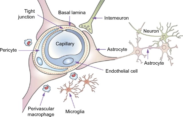
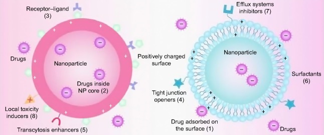
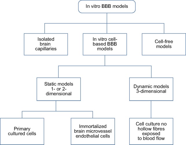
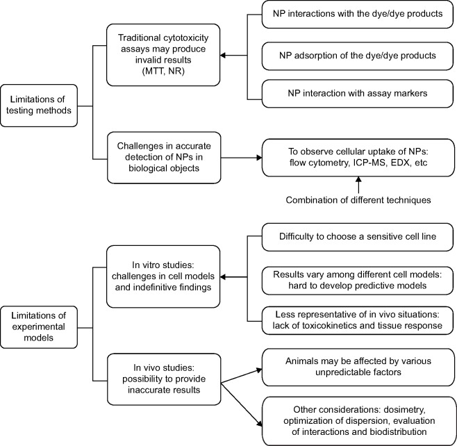
Nanomaterials are defined as materials with one or more external dimensions with a size of 1-100 nm. Such materials possess typical nanostructure-dependent properties (eg, chemical, biological, optical, mechanical, and magnetic), which may differ greatly from the properties of their bulk counterparts. In recent years, nanomaterials have been widely used in the production of dental materials, particularly in light polymerization composite resins and bonding systems, coating materials for dental implants, bioceramics, endodontic sealers, and mouthwashes. However, the dental applications of nanomaterials yield not only a significant improvement in clinical treatments but also growing concerns regarding their biosecurity. The brain is well protected by the blood-brain barrier (BBB), which separates the blood from the cerebral parenchyma. However, in recent years, many studies have found that nanoparticles (NPs), including nanocarriers, can transport through the BBB and locate in the central nervous system (CNS). Because the CNS may be a potential target organ of the nanomaterials, it is essential to determine the neurotoxic effects of NPs. In this review, possible dental nanomaterials and their pathways into the CNS are discussed, as well as related neurotoxicity effects underlying the in vitro and in vivo studies. Finally, we analyze the limitations of the current testing methods on the toxicological effects of nanomaterials. This review contributes to a better understanding of the nano-related risks to the CNS as well as the further development of safety assessment systems.

Keywords: central nervous system; dental; nanomaterials; risk assessment; testing methods; toxicity.


Figures

Figure 1
Figure 1
Schematic of the blood–brain barrier and the associated components of the neurovascular unit. Note: Reprinted from Adv Drug Deliv Rev, 64(7), Chen Y, Liu L. Modern methods for delivery of drugs across the blood–brain barrier. 640–665., Copyright (2012), with permission from Elsevier.
Figure 2
Figure 2
Schematic representation of multifunctional NPs as drug carriers across the BBB. Notes: Drugs can be adsorbed onto the surface of the NP due to the interactions between positive and negative charges (1) and may even be trapped inside its core (2). Different strategies have been applied for the transportation of drugs across the BBB: receptor–ligands for unique recognition and endocytosis (3); tight junction openers for improved intercellular penetration (4); transcytosis enhancers for the promotion of the transport of NP across the membranes (5); surfactants for the enhancement of membrane fluidization (6); efflux system inhibitors for the reduction of drug efflux (7); and local toxicity inducers for the increase in the permeability of the endothelial cells (8). Reproduced with permission of Informa Healthcare. Barbu E, Molnar E, Tsibouklis J, Gorecki DC. The potential for nanoparticle-based drug delivery to the brain: overcoming the blood–brain barrier. Expert Opin Drug Deliv. 6(6):553–565, copyright © 2009, Informa Healthcare. Abbreviations: NP, nanoparticle; BBB, blood–brain barrier.
Figure 3
Figure 3
G7 NP distribution in different brain regions and cell populations. Notes: Confocal microscopy images of brain cryosections in mice sacrificed 6 hours after an intraperitoneal injection of G7 NPs labeled with DAPI (blue), G7 NPs (red), and a number of antibodies (green) Low- (AD) and high-magnification (EH) images of the cerebral cortex (A and E), corpus callosum (B and F), choroid plexus (C and G), and subfornical organ (F and H). (EH) High-magnification images of the dashed squares indicated in (AD). (IL) High-magnification images showing single cells from the hippocampal formation. Scale bar =50 μm (AD) and 10 μm (EL). Reprinted from J Control Release, 174, Vilella A, Tosi G, Grabrucker AM, et al. Insight on the fate of CNS-targeted nanoparticles. Part I: Rab5-dependent cell-specific uptake and distribution. 195–201., Copyright (2014), with permission from Elsevier. Abbreviations: NP, nanoparticle; G7 NPs, glycopeptides-modified poly-lactide-co-glycolide NPs; DAPI, 4′,6-diamidino-2-phenylindole.
Figure 4
Figure 4
In vitro cell culture models for the studies on drug and NP transport through the BBB. Note: Reprinted from Adv Drug Deliv Rev, 64(7), Wong HL, Wu XY, Bendayan R. Nanotechnological advances for the delivery of CNS therapeutics. 686–700., Copyright (2012), with permission from Elsevier. Abbreviations: NP, nanoparticle; BBB, blood–brain barrier.
Figure 5
Figure 5
Existing problems in assessing the neurotoxicity of NPs. Note: This scheme summarizes the major limitations of the testing methods and experimental models. Abbreviations: NPs, nanoparticles; NR, neutral red; ICP-MS, induced coupled plasma mass spectroscopy; EDX, energy-dispersive X-ray spectroscopy; MTT, 3-(4,5-Dimethylthiazol-2-yl)-2,5-diphenyltetrazolium bromide.

Comment in

  • Potential toxicity of dental nanomaterials to the central nervous system.
    Solla DF, Paiva TS, André M, Paiva WS. Solla DF, et al. Int J Nanomedicine. 2015 Sep 3;10:5593-4. doi: 10.2147/IJN.S91856. eCollection 2015. Int J Nanomedicine. 2015. PMID: 26366079 Free PMC article. No abstract available.
  • Authors' reply.
    Feng X, Chen A, Zhang Y, Wang J, Shao L, Wei L. Feng X, et al. Int J Nanomedicine. 2015;10:5595-6. Int J Nanomedicine. 2015. PMID: 26594672 No abstract available.

References

    1. Bleeker EA, de Jong WH, Geertsma RE, et al. Considerations on the EU definition of a nanomaterial: science to support policy making. Regul Toxicol Pharm. 2013;65(1):119–125. - PubMed
    1. Juanola-Feliu E. The nanotechnology revolution in Barcelona: innovation and creativity by universities. Manage Int. 2009;13:111–123.
    1. Kasraei S, Sami L, Hendi S, Alikhani MY, Rezaei-Soufi L, Khamverdi Z. Antibacterial properties of composite resins incorporating silver and zinc oxide nanoparticles on Streptococcus mutans and Lactobacillus. Restor Dent Endod. 2014;39(2):109–114. - PMC - PubMed
    1. Niu LN, Fang M, Jiao K, et al. Tetrapod-like zinc oxide whisker enhancement of resin composite. J Dent Res. 2010;89(7):746–750. - PubMed
    1. Memarzadeh K, Sharili AS, Huang J, Rawlinson SC, Allaker RP. Nanoparticulate zinc oxide as a coating material for orthopedic and dental implants. J Biomed Mater Res A. 2015;103(3):981–989. - PubMed

Publication types