Review
. 2015 May 7:6:219.
doi: 10.3389/fimmu.2015.00219. eCollection 2015.

Shigella manipulates host immune responses by delivering effector proteins with specific roles

Affiliations
Review

Shigella manipulates host immune responses by delivering effector proteins with specific roles

Hiroshi Ashida et al. Front Immunol. .

Abstract

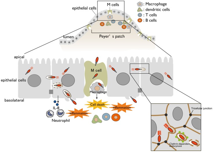
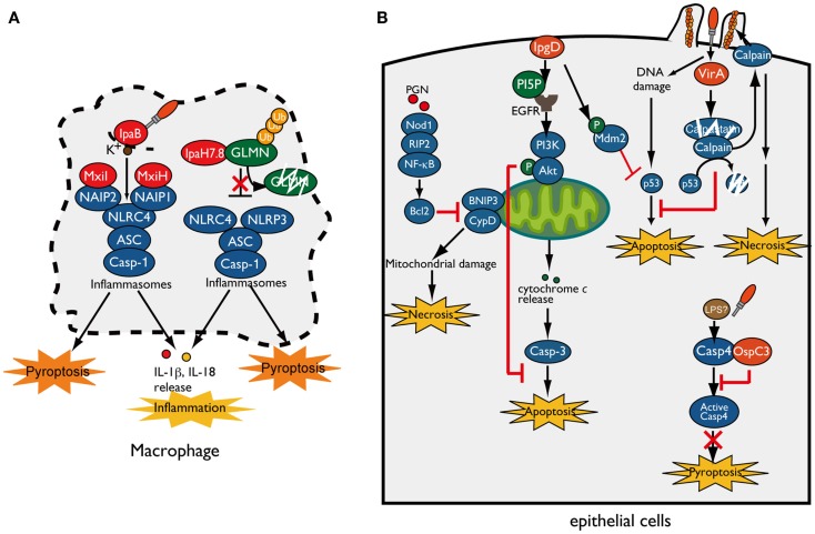
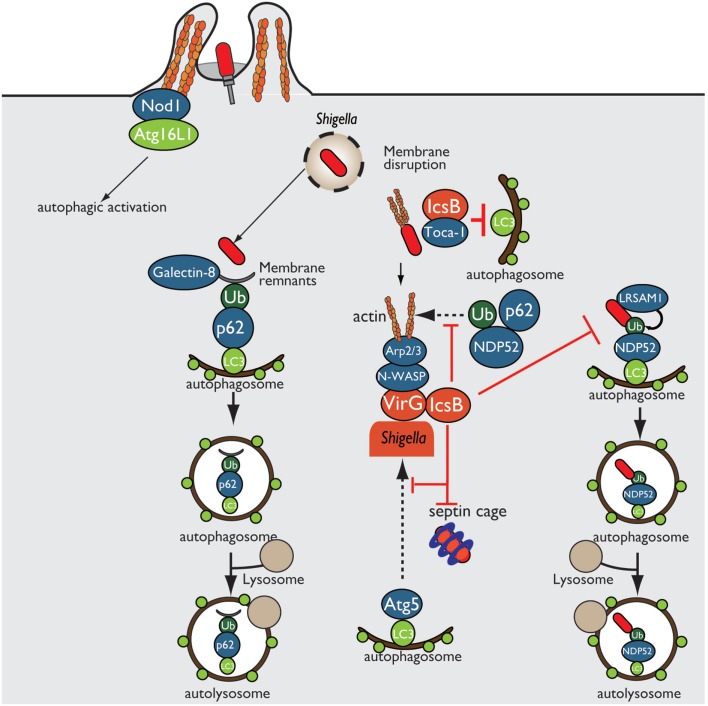
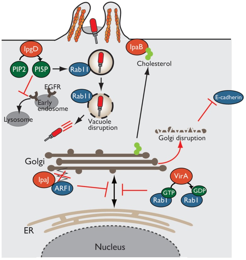
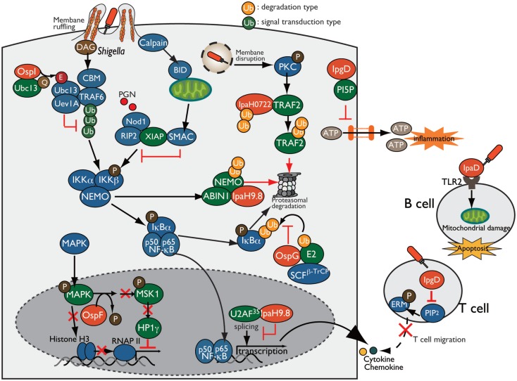
The intestinal epithelium deploys multiple defense systems against microbial infection to sense bacterial components and danger alarms, as well as to induce intracellular signal transduction cascades that trigger both the innate and the adaptive immune systems, which are pivotal for bacterial elimination. However, many enteric bacterial pathogens, including Shigella, deliver a subset of virulence proteins (effectors) via the type III secretion system (T3SS) that enable bacterial evasion from host immune systems; consequently, these pathogens are able to efficiently colonize the intestinal epithelium. In this review, we present and select recently discovered examples of interactions between Shigella and host immune responses, with particular emphasis on strategies that bacteria use to manipulate inflammatory outputs of host-cell responses such as cell death, membrane trafficking, and innate and adaptive immune responses.

Keywords: Shigella; effector; inflammation; innate immunity.


Figures

Figure 1
Figure 1
A model for Shigella infection of the intestinal epithelium. A schematic representation of Shigella infection. Bacterial invasion and multiplication within macrophages and epithelial cells cause subsequent massive inflammatory colitis, termed as shigellosis. Cell-to-cell movement of Shigella occurs at tricellular junctions via clathrin-dependent endocytosis.
Figure 2
Figure 2
Shigella manipulates host-cell death. (A) Shigella invasion and replication in macrophages trigger NLRC4/NLRP3-inflammasomes activation and pyroptotic cell death. The Shigella T3SS rod component MxiI and needle protein MxiH trigger NAIP2- and NAIP1-dependent NLRC4-inflammasome activation, respectively, whereas T3SS effector IpaB-mediated potassium influx triggers NLRC4-inflammasome activation and ultimately induces pyroptosis. IpaH7.8 ubiquitinates GLMN, and undergoes proteasome-dependent degradation, resulting in NLRP3/NLRC4-inflammasome activation and pyroptosis. (B) Shigella counteracts mitochondrial damage-dependent necrosis through BNIP3 and CypD by activating the PGN/Nod1/RIP2/NF-κB/Bcl2 pro-survival pathway. Furthermore, Shigella prevents epithelial cell death by delivering an array of T3SS effectors. PI5P, generated by IpgD, promotes EGFR activation, which contributes to sustained activation of the PI3K–Akt survival pathway. IpgD and VirA target Mdm2 and calpain, respectively, to prevent p53-dependent apoptosis. OspC3 binds to the caspase-4 subunit and inhibits its activation, thereby blocking caspase-4-dependent inflammatory cell death.
Figure 3
Figure 3
Shigella prevents autophagic recognition. Shigella prevents autophagic clearance by evading autophagic recognition. Shigella delivers the T3SS effector IcsB, which binds to the bacterial outer membrane protein VirG and prevents Atg5–VirG interaction and ubiquitin recruitment, thereby evading autophagic recognition. IcsB also recruits Toca-1, and prevents the recruitment of LC3 to around intracellular bacteria.
Figure 4
Figure 4
Shigella alters host intracellular trafficking. Shigella exploits and alters intracellular trafficking to manipulate the host defense system. PI5P produced by IpgD recruits Rab11 to the bacterial entry site and promotes vacuolar membrane disruption. Production of PI5P by IpgD further relocates EGFR in early endosomes, resulting in blocking lysosomal degradation of EGFR and sustaining PI3K–Akt survival pathway. VirA and IpaJ target and inactivate Rab1 and ARF1, respectively, thereby preventing intracellular trafficking and Golgi disruption. IpaB binds to and relocates cholesterol from Golgi to bacterial entry sites, resulting in Golgi fragmentation.
Figure 5
Figure 5
Shigella manipulates the host innate and adaptive immune response. Shigella manipulates host inflammatory responses by delivering a subset of T3SS effectors, including IpgD, OspI, OspG, OspF, and IpaH. PI5P, generated by IpgD, prevents ATP release-dependent inflammation through hemichannels. OspG binds to ubiquitin and ubiquitinated E2 proteins and prevents IκBα ubiquitylation, which is required for NF-κB activation. OspI deamidates and inactivates Ubc13, resulting in inhibition of TRAF6 ubiquitination. IpaH0722 and IpaH9.8 target TRAF2 and NEMO, respectively, for ubiquitination, and undergo proteasome degradation. In addition, IpaH9.8 further targets U2AF35, preventing its participation in the splicing reaction via an unidentified bacterial factor, Shigella induces calpain-dependent BID activation, which, in turn, releases mitochondrial SMAC to antagonize XIAP-mediated inflammation. IpgD-mediated hydrolysis of PIP2 inactivates ERMs of T cells, which crosslink actin filaments with the plasma membrane, thereby preventing T cell migration. Shigella induces B cell death in both invaded and non-invaded cells. In non-invaded cells, the T3SS needle-tip protein IpaD interacts with TLR2 on B cells and triggers mitochondrial damage, which eventually induces apoptosis.

References

    1. Parsot C. Shigella type III secretion effectors: how, where, when, for what purposes? Curr Opin Microbiol (2009) 12:110–6.10.1016/j.mib.2008.12.002 - DOI - PubMed
    1. Ashida H, Ogawa M, Mimuro H, Kobayashi T, Sanada T, Sasakawa C. Shigella are versatile mucosal pathogens that circumvent the host innate immune system. Curr Opin Immunol (2011) 23:448–55.10.1016/j.coi.2011.06.001 - DOI - PubMed
    1. Wassef JS, Keren DF, Mailloux JL. Role of M cells in initial antigen uptake and in ulcer formation in the rabbit intestinal loop model of shigellosis. Infect Immun (1989) 57:858–63. - PMC - PubMed
    1. Perdomo OJ, Cavaillon JM, Huerre M, Ohayon H, Gounon P, Sansonetti PJ. Acute inflammation causes epithelial invasion and mucosal destruction in experimental shigellosis. J Exp Med (1994) 180:1307–19.10.1084/jem.180.4.1307 - DOI - PMC - PubMed
    1. High N, Mounier J, Prévost MC, Sansonetti PJ. IpaB of Shigella flexneri causes entry into epithelial cells and escape from the phagocytic vacuole. EMBO J (1992) 11:1991–9. - PMC - PubMed

LinkOut - more resources