Review
. 2015 May 5:6:288.
doi: 10.3389/fpls.2015.00288. eCollection 2015.

NAC-MYB-based transcriptional regulation of secondary cell wall biosynthesis in land plants

Affiliations
Review

NAC-MYB-based transcriptional regulation of secondary cell wall biosynthesis in land plants

Yoshimi Nakano et al. Front Plant Sci. .

Abstract

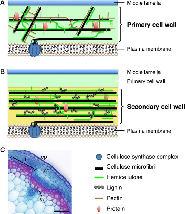
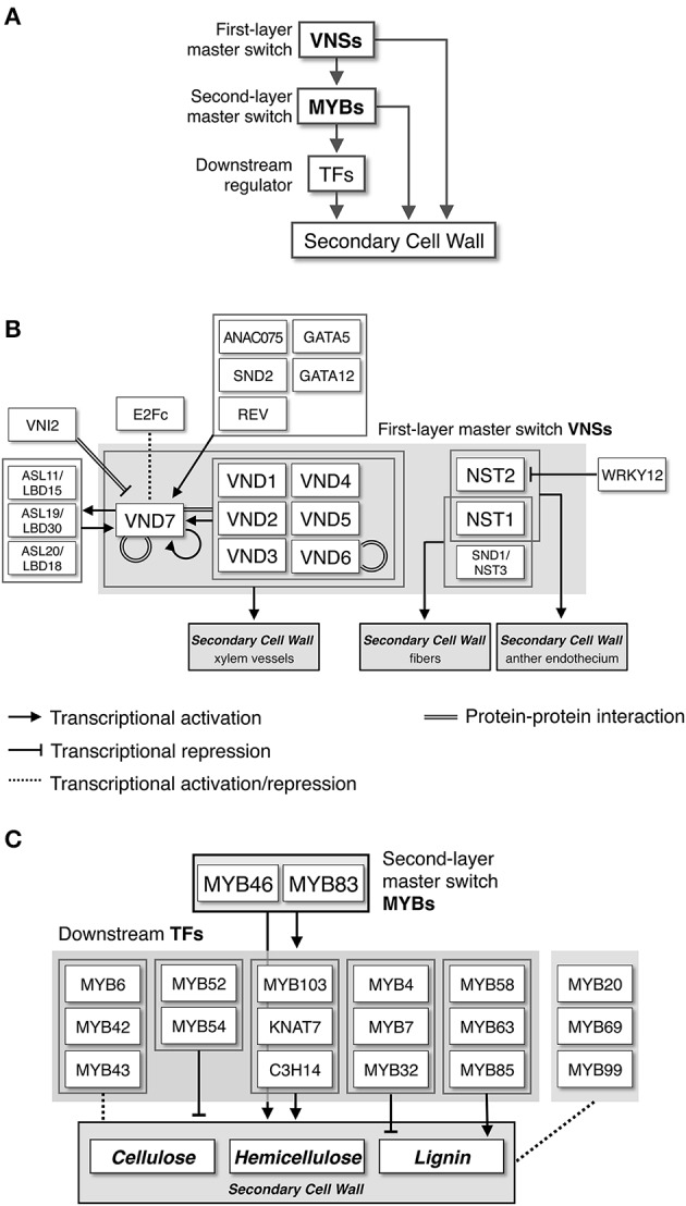
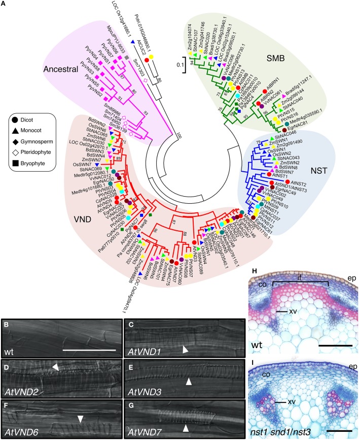
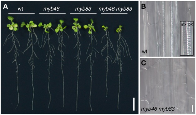
Plant cells biosynthesize primary cell walls (PCW) in all cells and produce secondary cell walls (SCWs) in specific cell types that conduct water and/or provide mechanical support, such as xylem vessels and fibers. The characteristic mechanical stiffness, chemical recalcitrance, and hydrophobic nature of SCWs result from the organization of SCW-specific biopolymers, i.e., highly ordered cellulose, hemicellulose, and lignin. Synthesis of these SCW-specific biopolymers requires SCW-specific enzymes that are regulated by SCW-specific transcription factors. In this review, we summarize our current knowledge of the transcriptional regulation of SCW formation in plant cells. Advances in research on SCW biosynthesis during the past decade have expanded our understanding of the transcriptional regulation of SCW formation, particularly the functions of the NAC and MYB transcription factors. Focusing on the NAC-MYB-based transcriptional network, we discuss the regulatory systems that evolved in land plants to modify the cell wall to serve as a key component of structures that conduct water and provide mechanical support.

Keywords: MYB transcription factor; NAC transcription factor; land plant evolution; network; secondary cell wall.


Figures

Figure 1
Figure 1
Plant cell walls. (A) Model of the primary cell wall. Cellulose microfibrils in the primary cell wall are relatively short and thin, compared with those in the secondary cell wall, and hemicellulose in the primary cell wall is composed of xyloglucan. The primary cell wall is rich in pectin. (B) Model of the secondary cell wall, which is deposited between the primary cell wall and the plasma membrane. The secondary cell wall mainly contains relatively long and thick cellulose microfibrils, hemicellulosic xylan, and lignin. (C) Cross section of an Arabidopsis inflorescence stem stained with Safranin, which stains lignin red, and Astra blue. co, cortex; ep, epidermis; if, interfascicular fiber; xv, xylem vessel. Bar = 50 μm.
Figure 2
Figure 2
Transcriptional network regulating secondary cell wall formation. (A) NAC-MYB-based transcriptional regulation of secondary cell wall biosynthesis. Some metabolic genes for secondary cell wall biosynthesis are targeted by both NACs and MYBs, producing “feed-forward” regulation. (B) Transcriptional regulatory network around VNS proteins, first-layer master switches for secondary cell wall formation, based on work in Arabidopsis. (C) Transcriptional regulatory network around MYB proteins, second-layer master switches for secondary cell wall formation, based on work in Arabidopsis.
Figure 3
Figure 3
The VNS genes function as first-layer master switches for secondary cell wall formation. (A) Phylogenetic tree of VNS proteins. The unrooted phylogenetic tree was constructed with amino acid sequences of the NAC domain (sequences provided in Table S1) by the maximum-likelihood method. Numbers indicate bootstrap values for the clades that received support values of over 70% (1000 resamplings). Scale (0.1) represents a 10% change in sequences. Based on the tree, the VNS proteins are classified into four groups, VND, NST/SND, SMB, and Ancestral groups. (B–G) Seven-day-old Arabidopsis roots of wild type (wt, B) and transgenic plants, in which AtVND1 (C), AtVND2 (D), AtVND3 (E), AtVND6 (F), and AtVND7 (G) were overexpressed by an inducible system. Ectopic xylem elements formed in the transgenic roots (white arrowheads). Data were adapted from Endo et al. (2015). (H,I) Cross sections of Arabidopsis inflorescence stems stained with Safranin, which stains lignin red, and Astra blue. In the wild type (wt), both xylem vessel cells and interfascicular fiber cells have lignin-containing secondary cell wall, thus they stain red (H). By contrast, the mutant nst1 snd1/nst3 lacks secondary cell wall in interfascicular fiber cells, thus only xylem vessel cells were stained by Safranin (I), as described in Mitsuda et al. (2007). co, cortex; ep, epidermis; if, interfascicular fiber; xv, xylem vessel. Bars = 100 μm (B–I).
Figure 4
Figure 4
The MYB46/83 genes function as second-layer master switches for secondary cell wall formation. (A) Ten-day-old Arabidopsis seedlings of wild type (wt), myb46, myb83, and myb46 myb83 mutants. The myb46 myb83 mutant shows growth inhibition in aerial parts. (B,C) Xylem vessels in the roots of the wild type (wt, B) and myb46 myb83 mutants (C). In the wild type, thick secondary cell wall is deposited in protoxylem-type (px) and metaxylem-type (mx) vessel cells (inset in B). In the myb46 myb83 mutant, secondary cell wall deposition in xylem vessel cells is strongly inhibited, as described in McCarthy et al. (2009). Bars = 1 cm (A) and 25 μm (B,C).
Figure 5
Figure 5
The PpVNS genes function in cell wall thickening in the moss P. patens. Stereid cells in leaf vein of the wild type (A) and ppvns1 ppvns6 pvns7 triple mutant (B). In the triple mutants, the stereid cell walls were significantly less thick, suggesting the importance of PpVNS proteins in cell wall thickening in the moss. Data were adapted from Xu et al. (2014). Bar = 5 μm.

References

    1. Allona I., Quinn M., Shoop E., Swope K., St Cyr S., Carlis J., et al. (1998). Analysis of xylem formation in pine by cDNA sequencing. Proc. Natl. Acad. Sci. U.S.A. 95, 9693–9698. 10.1073/pnas.95.16.9693 - DOI - PMC - PubMed
    1. Becker-André M., Schulze-Lefert P., Hahlbrock K. (1991). Structural comparison, modes of expression, and putative cis-acting elements of the two 4-coumarate: CoA ligase genes in potato. J. Biol. Chem. 266, 8551–8559. - PubMed
    1. Bedon F., Grima-Pettenati J., Mackay J. (2007). Conifer R2R3-MYB transcriotion factors: sequence analyses and gene expression in wood-forming tissues of white spruce (Picea glauca). BMC Plant Biol. 7:17. 10.1186/1471-2229-7-17 - DOI - PMC - PubMed
    1. Bell-Lelong D. A., Cusumano J. C., Meyer K., Chapple C. (1997). Cinnamate-4-hydroxylase expression in Arabidopsis. Regulation in response to development and the environment. Plant Physiol. 113, 729–738. 10.1104/pp.113.3.729 - DOI - PMC - PubMed
    1. Bennett T., van den Toorn A., Sanchez-Perez G. F., Campiho A., Willemsen V., Snel B., et al. (2010). SOMBRERO, BEARSKIN1, and BEARSKIN2 regulate root cap maturation in Arabidopsis. Plant Cell 22, 640–654. 10.1105/tpc.109.072272 - DOI - PMC - PubMed