Review
. 2015 May 5:6:306.
doi: 10.3389/fpls.2015.00306. eCollection 2015.

Information flow and protein dynamics: the interplay between nuclear magnetic resonance spectroscopy and molecular dynamics simulations

Affiliations
Review

Information flow and protein dynamics: the interplay between nuclear magnetic resonance spectroscopy and molecular dynamics simulations

Nina Pastor et al. Front Plant Sci. .

Abstract

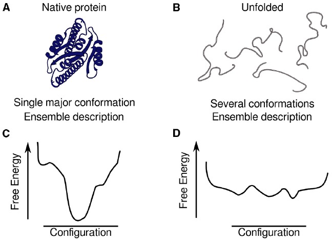
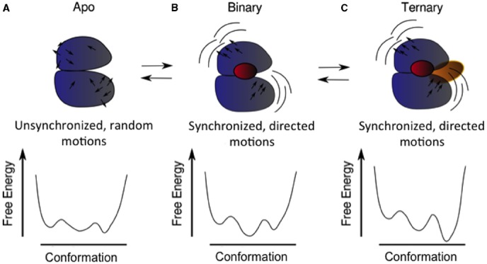
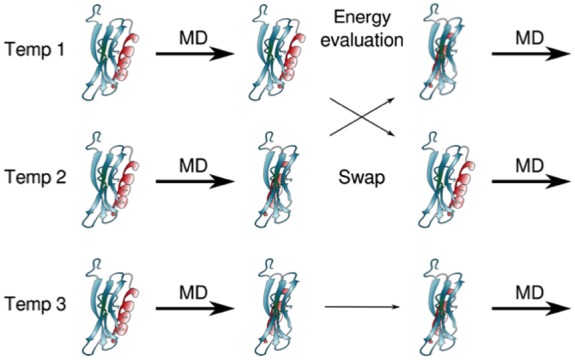
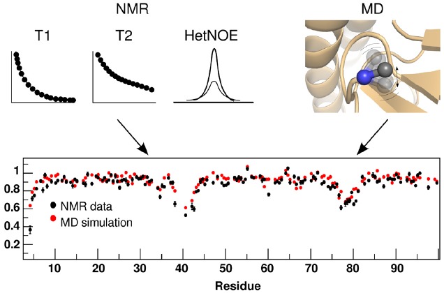
Proteins participate in information pathways in cells, both as links in the chain of signals, and as the ultimate effectors. Upon ligand binding, proteins undergo conformation and motion changes, which can be sensed by the following link in the chain of information. Nuclear magnetic resonance (NMR) spectroscopy and molecular dynamics (MD) simulations represent powerful tools for examining the time-dependent function of biological molecules. The recent advances in NMR and the availability of faster computers have opened the door to more detailed analyses of structure, dynamics, and interactions. Here we briefly describe the recent applications that allow NMR spectroscopy and MD simulations to offer unique insight into the basic motions that underlie information transfer within and between cells.

Keywords: aggregation; allosterism; binding; dynamics; folding intermediates.


Figures

FIGURE 1
FIGURE 1
Depiction of the ensembles of structures by conformational landscapes, with the location of minima, their depths and the heights of the barriers. Schematic representation of ensembles of structures for a globular, natively folded protein (A) and an intrinsically disordered protein (B). The description of a globular protein is based on an ensemble of structures clustered around a deep global minimum that can have substates in it (C) but represent basically the same topology, whereas the description of an IDP is based on a dynamic ensemble of different conformations represented by a collection of minima with similar depths (D).
FIGURE 2
FIGURE 2
Schematic of structural and dynamic coupling between different functional sites in a protein and how this coupling could modulate the energy landscape. The apo or ligand-free protein (A) samples three conformations in its native basin and displays unsynchronized and undirected motions (black arrows). Upon binding of the first ligand (B), a particular conformation is selected (the minimum in the middle of the landscape), and also the motions of the two main lobules of the protein become synchronized (notice the similar direction of the arrows in each domain). These motions favor the binding of the second ligand (C), selecting another of the possible conformations.
FIGURE 3
FIGURE 3
Example of an enhanced sampling method consisting on multiple MD simulation replicas at different temperatures (each row). At defined time intervals, which can vary typically from a few to hundreds of ps, the energy of the structures of each replica are evaluated. If the energy of run 1 is lower than that of run 2, or if it is higher but allowed by the Boltzmann factor, the replicas are exchanged, so now the structure from run 1 is simulated at temperature 2, and the structure from run 2 is simulated at temperature 1 (exchange between the top and middle rows). Otherwise, as shown for temperature 3 (lower row), there is no exchange and run 3 continues to be simulated at temperature 3. Exchanges are attempted until all replicas have been run at all temperatures. Structures are then collected as a function of temperature, and data can be analyzed for all temperatures, or only for a particular temperature, of functional or physiological relevance.
FIGURE 4
FIGURE 4
Correspondence of dynamic parameters measured by NMR and back-calculated from an MD trajectory. The square of the backbone order parameter (S2 shown in the bottom panel), which provides information about the amplitude of the motion of NH bonds or CH bonds, can be calculated from measured experimental parameters T1, T2, and HetNOE (black dots in the bottom panel), and from the MD trajectories (red dots in the bottom panel). The resulting values, for both approaches, can be compared directly.

References

    1. Aglietti R. A., Floor S. N., McClendon C. L., Jacobson M. P., Gross J. D. (2013). Active site conformational dynamics are coupled to catalysis in the mRNA decapping enzyme Dcp2. Structure 21, 1571–1580. 10.1016/j.str.2013.06.021 - DOI - PMC - PubMed
    1. Allison J. R. (2012). Assessing and refining molecular dynamics simulations of proteins with nuclear magnetic resonance data. Biophys. Rev. 4, 189–203 10.1007/s12551-012-0087-6 - DOI - PMC - PubMed
    1. Allison J. R., Hertig S., Missimer J. H., Smith L. J., Steinmetz M. O., Dolenc J. (2012). Probing the structure and dynamics of proteins by combining molecular dynamics simulations and experimental NMR data. J. Chem. Theory Comput. 8, 3430–3444 10.1021/ct300393b - DOI - PubMed
    1. Allnér O., Foloppe N., Nilsson L. (2015). Motions and entropies in proteins as seen in NMR relaxation experiments and molecular dynamics simulations. J. Phys. Chem. B 119, 1114–1128. 10.1021/jp506609g - DOI - PubMed
    1. Baker C. M., Best R. B. (2014). Insights into the binding of intrinsically disordered proteins from molecular dynamics simulation. Wiley Interdiscip. Rev. Comput. Mol. Sci. 4, 182–198 10.1002/wcms.1167 - DOI - PMC - PubMed

LinkOut - more resources