

The viability of mouse spermatogonial germ cells on a novel scaffold, containing human serum albumin and calcium phosphate nanoparticles
- PMID: 26000004
- PMCID: PMC4426153
The viability of mouse spermatogonial germ cells on a novel scaffold, containing human serum albumin and calcium phosphate nanoparticles
Abstract
Background: In spermatogenesis, spermatogonial cells differentiate to the haploid gametes. It has been shown that spermatogenesis can be done at in vitro condition. In vitro spermatogenesis may provide an open window to treat male infertility.
Objective: The aim of this study was to evaluate the effects of a novel scaffold containing human serum albumin (HSA)/tri calcium phosphate nanoparticles (TCP NPs) on the mouse spermatogonial cell line (SCL).
Materials and methods: First, TCP NPs were synthesized by reaction of calcium nitrate and diammonium phosphate at pH 13. Then, serial concentrations of TCP NPs were separately added to 500 mg/mL HSA, and incubated in the 100(o)C water for 30 min. In the next step, each scaffold was cut (2×2mm), placed into sterile well of microplate, and then incubated for 1, 2, and 3 days at 37(o)C with mouse SCL. After incubation, the cytotoxicity of the scaffolds was evaluated by different tests including 3-(4,5-dimethylthiazol-2-yl)-2, 5-diphenyl-tetrazolium bromide (MTT) assay, lactate dehydrogenase (LDH) assay, vital staining, and cell counting. On the other hand, the release of TCP NPs and HSA from the scaffolds was measured.
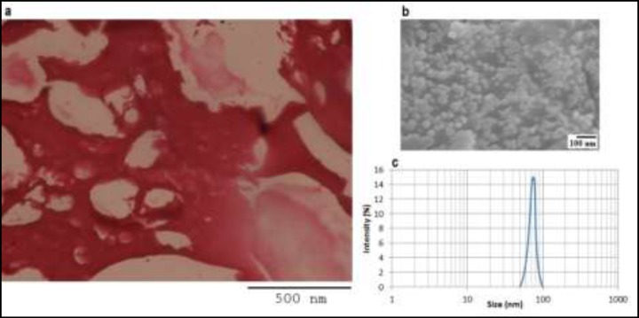
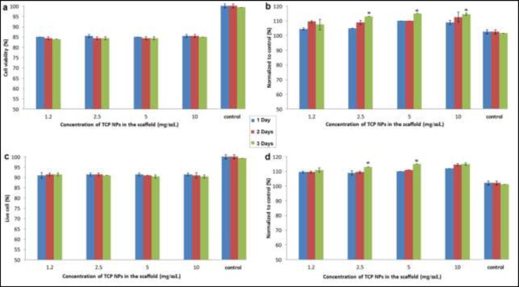
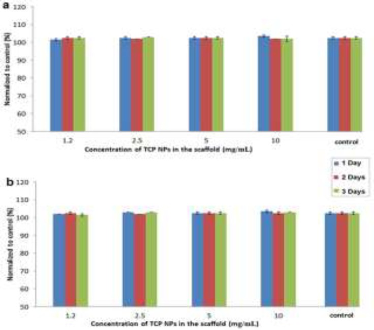
Results: Based on microscopic observation, the size of cavities for all scaffolds was near 200-500 µm, and the size of TCP NPs was near 50-100 nm. All toxicity tests showed that the increase of TCP concentration in the scaffold did not affect mouse SCL. It means that the percentage of cell viability, LDH release, vital cells, and cell quantity was 85%, 105%, 90%, and 110%, respectively. But, the increase of incubation time led to increase of LDH release (up to 115%) and cell count (up to 115%). Also, little decrease of cell viability and vital cells was seen when incubation time was increased. Here, no release of TCP NPs and HSA was seen after increase of TCP concentration and incubation time.
Conclusion: It can be concluded that the increase of TCP concentration in HSA/ TCP NPs scaffold does not lead to cytotoxicity. On the other hand, the increase of incubation time leads to increase of mouse SCL cell death. In this study, it was found that TCP NPs and HSA could not release from the scaffolds. In future, both proliferation and differentiation of mouse SCL on HSA/TCP NPs scaffold must be checked over more wide incubation times.
Keywords: Calcium phosphate nanoparticles; Cytotoxic effect; Human serum albumin; Scaffold; Spermatogonial cell line.
Figures
Similar articles
-
The Viability of Human Testis-Derived Cells on Human Serum Albumin-Based Scaffold as An Artificial Male Germ Cell Niche.Int J Fertil Steril. 2020 Jul;14(2):150-153. doi: 10.22074/ijfs.2020.6086. Epub 2020 Jul 15. Int J Fertil Steril. 2020. PMID: 32681628 Free PMC article.
-
Surface modification of tri-calcium phosphate nanoparticles by DOPE and/or anti-E6 antibody to enhance uptake of antisense of E6 mRNA.Colloids Surf B Biointerfaces. 2015 Feb 1;126:297-302. doi: 10.1016/j.colsurfb.2014.12.040. Epub 2015 Jan 3. Colloids Surf B Biointerfaces. 2015. PMID: 25601794
-
Structural and degradation characteristics of an innovative porous PLGA/TCP scaffold incorporated with bioactive molecular icaritin.Biomed Mater. 2010 Oct;5(5):054109. doi: 10.1088/1748-6041/5/5/054109. Epub 2010 Sep 28. Biomed Mater. 2010. PMID: 20876954
-
Human Serum Albumin Nanoparticles for Use in Cancer Drug Delivery: Process Optimization and In Vitro Characterization.Nanomaterials (Basel). 2016 Jun 15;6(6):116. doi: 10.3390/nano6060116. Nanomaterials (Basel). 2016. PMID: 28335244 Free PMC article.
-
Future of Spermatogonial Stem Cell Culture: Application of Nanofiber Scaffolds.Curr Stem Cell Res Ther. 2017;12(7):544-553. doi: 10.2174/1574888X12666170623095457. Curr Stem Cell Res Ther. 2017. PMID: 28641554 Review.
Cited by
-
Biomaterials for Testicular Bioengineering: How far have we come and where do we have to go?Front Endocrinol (Lausanne). 2023 Mar 16;14:1085872. doi: 10.3389/fendo.2023.1085872. eCollection 2023. Front Endocrinol (Lausanne). 2023. PMID: 37008920 Free PMC article. Review.
-
Advanced bioengineering of male germ stem cells to preserve fertility.J Tissue Eng. 2021 Nov 29;12:20417314211060590. doi: 10.1177/20417314211060590. eCollection 2021 Jan-Dec. J Tissue Eng. 2021. PMID: 34868541 Free PMC article. Review.
-
The Viability of Human Testis-Derived Cells on Human Serum Albumin-Based Scaffold as An Artificial Male Germ Cell Niche.Int J Fertil Steril. 2020 Jul;14(2):150-153. doi: 10.22074/ijfs.2020.6086. Epub 2020 Jul 15. Int J Fertil Steril. 2020. PMID: 32681628 Free PMC article.
References
LinkOut - more resources
Full Text Sources
Research Materials
Miscellaneous