. 2015 May;2(5):518-33.
doi: 10.1002/acn3.193. Epub 2015 Mar 27.

Sulfatide levels correlate with severity of neuropathy in metachromatic leukodystrophy

Affiliations

Sulfatide levels correlate with severity of neuropathy in metachromatic leukodystrophy

Christine Í Dali et al. Ann Clin Transl Neurol. 2015 May.

Abstract

Objective: Metachromatic leukodystrophy (MLD) is an autosomal recessive lysosomal storage disorder due to deficient activity of arylsulfatase A (ASA) that causes accumulation of sulfatide and lysosulfatide. The disorder is associated with demyelination and axonal loss in the central and peripheral nervous systems. The late infantile form has an early-onset, rapidly progressive course with severe sensorimotor dysfunction. The relationship between the degree of nerve damage and (lyso)sulfatide accumulation is, however, not established.

Methods: In 13 children aged 2-5 years with severe motor impairment, markedly elevated cerebrospinal fluid (CSF) and sural nerve sulfatide and lysosulfatide levels, genotype, ASA mRNA levels, residual ASA, and protein cross-reactive immunological material (CRIM) confirmed the diagnosis. We studied the relationship between (lyso)sulfatide levels and (1) the clinical deficit in gross motor function (GMFM-88), (2) median and peroneal nerve motor and median and sural nerve sensory conduction studies (NCS), (3) median and tibial nerve somatosensory evoked potentials (SSEPs), (4) sural nerve histopathology, and (5) brain MR spectroscopy.

Results: Eleven patients had a sensory-motor demyelinating neuropathy on electrophysiological testing, whereas two patients had normal studies. Sural nerve and CSF (lyso)sulfatide levels strongly correlated with abnormalities in electrophysiological parameters and large myelinated fiber loss in the sural nerve, but there were no associations between (lyso)sulfatide levels and measures of central nervous system (CNS) involvement (GMFM-88 score, SSEP, and MR spectroscopy).

Interpretation: Nerve and CSF sulfatide and lysosulfatide accumulation provides a marker of disease severity in the PNS only; it does not reflect the extent of CNS involvement by the disease process. The magnitude of the biochemical disturbance produces a continuously graded spectrum of impairments in neurophysiological function and sural nerve histopathology.


Figures

Figure 1
Figure 1
Composite of light microscopy semithin sections stained with toluidine blue (A, arrows indicate metachromatic granules) and p-phenylenediamine (B) and ultrathin sections (E) from the right sural nerve of a 2.5-year-old girl with late infantile metachromatic leukodystrophy (MLD) (patient 1). The diameter distribution of myelinated fibers and the calculated total number from light microscopy are shown below left (D). The myelin versus fiber diameter relationship (F) and the g-ratio distribution (G) were obtained from the electron micrographs. The morphological data were compared to the nerve conduction studies of the same sural nerve (C).
Figure 2
Figure 2
Motor and sensory nerve conduction studies from the right median nerve (A and B, left column) and motor conduction studies from the right peroneal nerve (C and D, right column). The patient on top (A and C) was a 5-year-old boy (patient 8) without electrophysiological evidence of neuropathy, and the patient below (B and D) was a 3-year-old boy with severe demyelinating neuropathy (patient 4). The compound sensory nerve action potential (SNAP) of the median nerve was evoked by stimulation at Digit2 (Dig2) and recording at the wrist (Wr), the compound muscle action potential (CMAP) of the median nerve was evoked by stimulation at wrist (Wr) and elbow (Elb) and recorded at the abductor pollicis brevis muscle (APB). The CMAP of the peroneal nerve was evoked by stimulation at the ankle and distal to the fibular head (DCF) and recorded from the extensor digitorum brevis muscle (EDB). Latencies, conduction velocities, and amplitudes of SNAPs and CMAPs are indicated below traces.
Figure 3
Figure 3
Comparison of morphological (A and B) and electrophysiological (C) findings of the right sural nerves in three patients with late infantile metachromatic leukodystrophy (MLD). The three patients had either no evidence of neuropathy (top, 3-year-old girl, patient 7), moderate neuropathy (above middle, 4-year-old girl, patient 2), or severe neuropathy (below middle, 4-year-old boy, patient 11). The lowermost section was from a 3-year-old control without evidence of neuropathy. The diameter distribution and total number of fibers (B, center column) were calculated from light microscopy of p-phenyldiamine semithin sections. Sural nerve conduction studies are shown in the right column (C). The amplitudes and conduction velocities are indicated above the traces.
Figure 4
Figure 4
Cumulative distributions of g-ratios from sural nerves in 12 patients with late infantile metachromatic leukodystrophy (full lines) and three children without neuropathy (stippled lines).
Figure 5
Figure 5
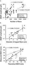
Relationships between sensory nerve action potentials (SNAP) and sural nerve myelinated fibers in patients with metachromatic leukodystrophy. (A) The SNAP amplitudes from the median nerve, the left sural nerve, and right sural nerves were correlated with the number of fibers with diameters above 9 μm (filled symbols, P < 0.001) and above 7 μm (open symbols, P < 0.001) in the right sural nerve. (B) The SNAP amplitudes in the right median nerve, left sural nerve, and right sural nerve were correlated with the mean diameters of the 10 largest fibers in the right sural nerve (P < 0.001). (C) Correlation between the measured sensory nerve conduction velocities (SNCV) in the right median nerve, left sural nerve, and right sural nerve and the theoretical SNCV calculated from the mean diameters of the 10 largest fibers in the right sural nerve using a conversion factor of 4.3 m/sec per μm. The stippled line indicates the predicted conduction velocity. The lower SNCV in the patients (solid line; P < 0.001) is consistent with demyelination.
Figure 6
Figure 6
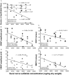
Relationships between sural nerve sulfatide concentration and myelinated fibers and nerve conduction studies in late infantile metachromatic leukodystrophy. (A) Number of myelinated fibers in the right sural nerve (fibers >9 μm, P < 0.05; fibers >7 μm, P = 0.07; all fibers, P = 0.9). (B) Pooled amplitudes of sensory nerve action potentials (SNAP) in the right median, left sural, and right sural nerves. (C) Pooled sensory nerve conduction velocities (SNCV) in the right median, left sural, and right sural nerves. (D) Pooled amplitudes of compound muscle action potentials (CMAP) in the right median and peroneal nerves. (E) Pooled motor nerve conduction velocities (MNCV) in the right median and peroneal nerves.
Figure 7
Figure 7
Relationship between the cerebrospinal fluid sulfatide concentration and median nerve somatosensory evoked potentials (SSEP). The latencies of the cortical responses correlated with the sulfatide concentration (P < 0.01) and the peripheral latencies trended toward significance (P = 0.06). The central conduction time was not correlated with the sulfatide concentration.

References

    1. Gieselmann V. Metachromatic leukodystrophy: genetics, pathogenesis and therapeutic options. Acta Paediatr Suppl. 2008;97:15–21. - PubMed
    1. Biffi A, Cesani M, Fumagalli F, et al. Metachromatic leukodystrophy – mutation analysis provides further evidence of genotype-phenotype correlation. Clin Genet. 2008;74:349–357. - PubMed
    1. Miller RG, Gutmann L, Lewis RA, Sumner AJ. Acquired versus familial demyelinative neuropathies in children. Muscle Nerve. 1985;8:205–210. - PubMed
    1. Zafeiriou DI, Kontopoulos EE, Michelakakis HM, et al. Neurophysiology and MRI in late-infantile metachromatic leukodystrophy. Pediatr Neurol. 1999;21:843–846. - PubMed
    1. Toda K, Kobayashi T, Goto I, et al. Lysosulfatide (sulfogalactosylsphingosine) accumulation in tissues from patients with metachromatic leukodystrophy. J Neurochem. 1990;55:1585–1591. - PubMed