

Primary Pulmonary NUT Midline Carcinoma: Clinical, Radiographic, and Pathologic Characterizations
- PMID: 26001144
- PMCID: PMC4443847
- DOI: 10.1097/JTO.0000000000000545
Primary Pulmonary NUT Midline Carcinoma: Clinical, Radiographic, and Pathologic Characterizations
Abstract
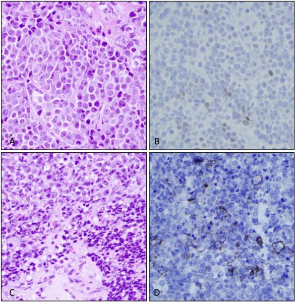
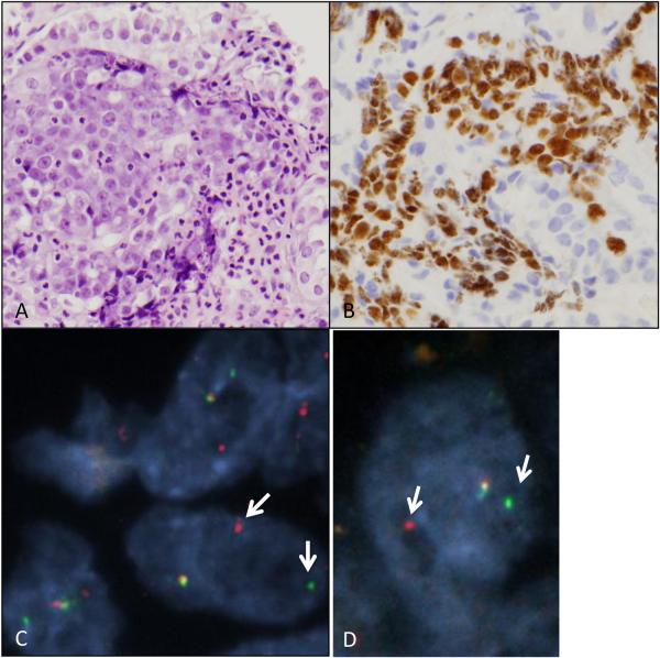
NUT midline carcinoma (NMC) is a poorly differentiated tumor typically driven by a t(15;19) rearrangement leading to a NUT fusion event. This rare and uniformly fatal tumor arises in multiple organ sites; however the clinical, radiographic, and pathologic characteristics of primary pulmonary NMC are poorly defined. We identified eight cases of primary pulmonary NMC in our consult practice over 4 years and, using a NUT immunohistochemistry screen, retrospectively identified one additional case from 166 (0.6%) consecutive in-house biopsies of lung carcinomas lacking glandular differentiation. Eight cases had available clinical and radiographic data and shared a remarkable degree of similarity. The median age at presentation was 30 (range 21-68). Six patients had little or no smoking history. All complained of 1 to 3 months of cough at presentation. Computed tomography scans showed a large, centrally located primary mass with confluent involvement of mediastinal lymph nodes, pleural disease, and sparing of the contralateral lung. Lytic bone metastases were common but brain metastases were absent in all cases. Pathologically, all cases showed primitive-appearing round to epitheloid cells growing in nests and sheets. All tumors expressed keratin, p63 or p40, and NUT protein. Eight cases had a fluorescence in situ hybridization-proven BRD4-NUT or BRD3-NUT rearrangement; one case was presumed to have a NUT-variant fusion event. Median overall survival was 2.2 months. Despite the rarity of primary pulmonary NMC, it is important to recognize this entity to counsel patients regarding outcome and to identify candidates for targeted BRD inhibitors currently in clinical trials.
Figures

References
-
- French CA. Pathogenesis of NUT midline carcinoma. Annu Rev Pathol. 2012;7:247–265. - PubMed
-
- French CA, Miyoshi I, Kubonishi I, et al. BRD4-NUT fusion oncogene: a novel mechanism in aggressive carcinoma. Cancer Res. 2003;63:304–307. - PubMed
-
- French CA, Ramirez CL, Kolmakova J, et al. BRD-NUT oncoproteins: a family of closely related nuclear proteins that block epithelial differentiation and maintain the growth of carcinoma cells. Oncogene. 2008;27:2237–2242. - PubMed
MeSH terms
Substances
Grants and funding
LinkOut - more resources
Full Text Sources
Medical
