. 2016 Jan;23(1):52-63.
doi: 10.1038/cdd.2015.66. Epub 2015 May 22.

Fyn-phosphorylated PIKE-A binds and inhibits AMPK signaling, blocking its tumor suppressive activity

Affiliations

Fyn-phosphorylated PIKE-A binds and inhibits AMPK signaling, blocking its tumor suppressive activity

S Zhang et al. Cell Death Differ. 2016 Jan.

Abstract

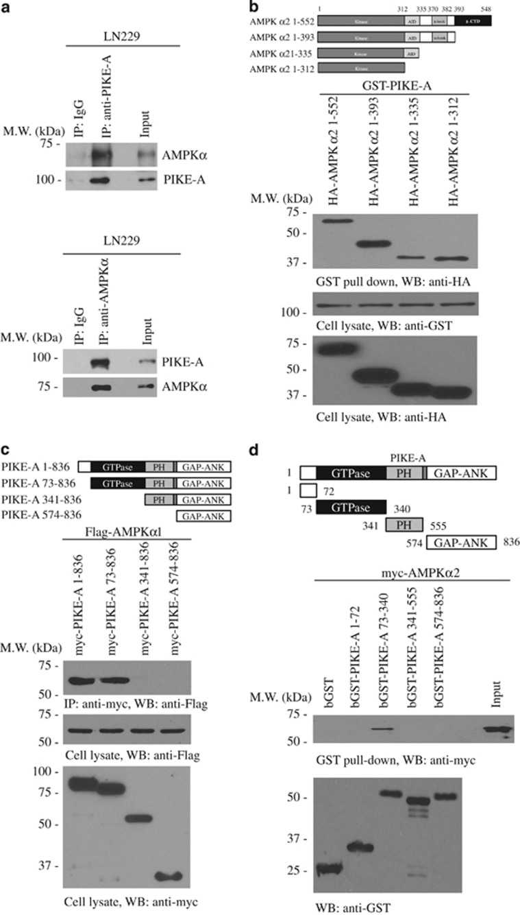
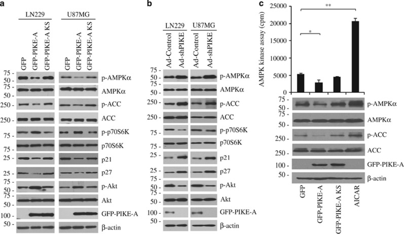
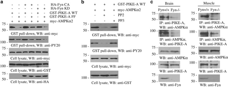
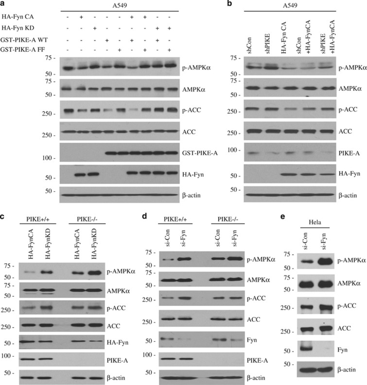
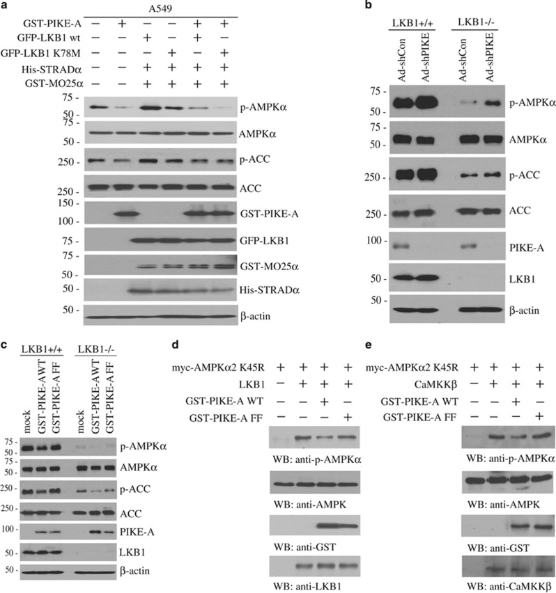
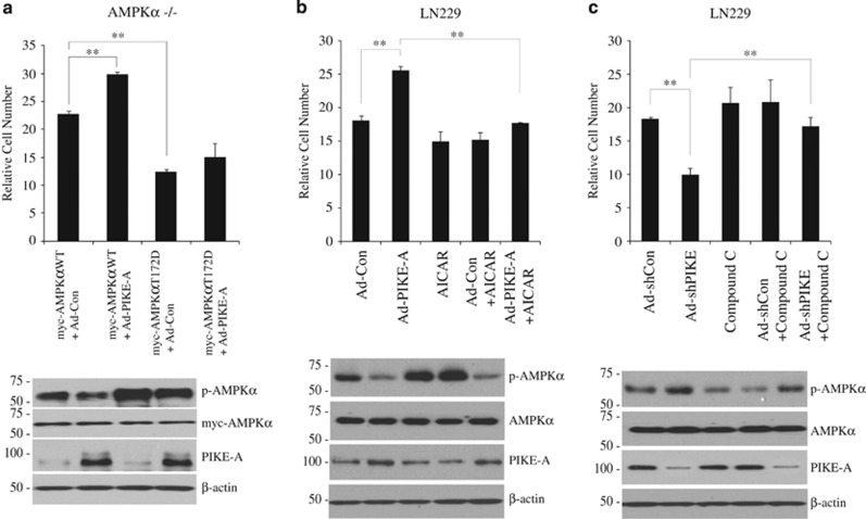
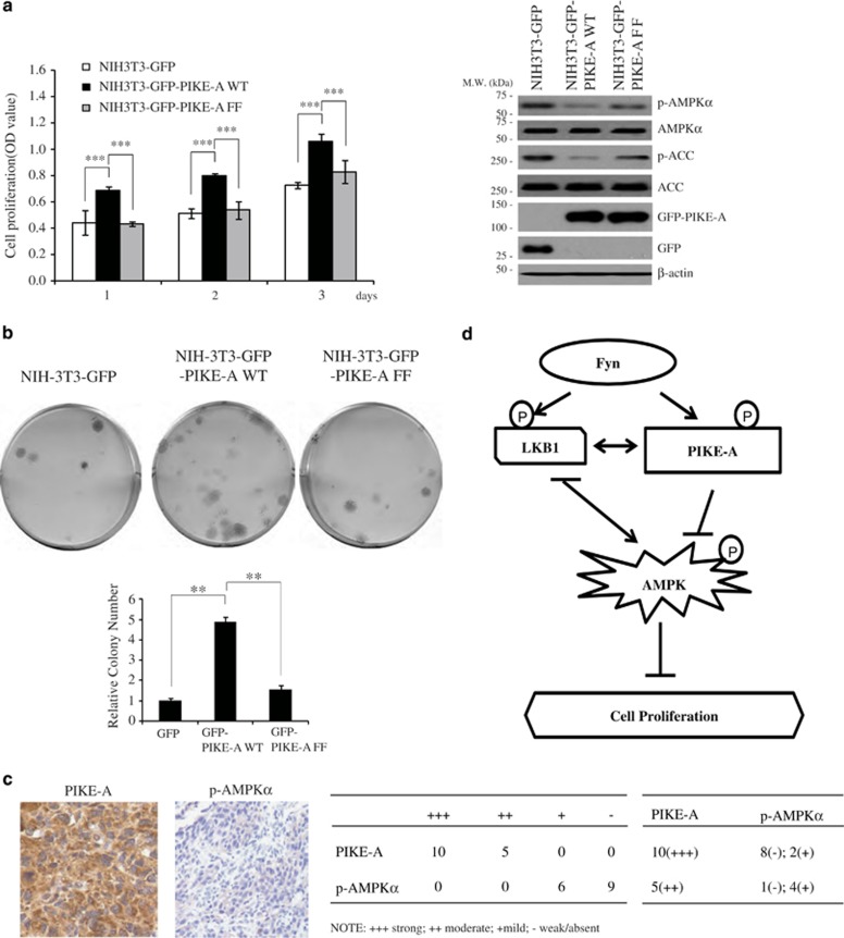
The AMP-activated protein kinase, a key regulator of energy homeostasis, has a critical role in metabolic disorders and cancers. AMPK is mainly regulated by cellular AMP and phosphorylation by upstream kinases. Here, we show that PIKE-A binds to AMPK and blocks its tumor suppressive actions, which are mediated by tyrosine kinase Fyn. PIKE-A directly interacts with AMPK catalytic alpha subunit and impairs T172 phosphorylation, leading to repression of its kinase activity on the downstream targets. Mutation of Fyn phosphorylation sites on PIKE-A, depletion of Fyn, or pharmacological inhibition of Fyn blunts the association between PIKE-A and AMPK, resulting in loss of its inhibitory effect on AMPK. Cell proliferation and oncogenic assays demonstrate that PIKE-A antagonizes tumor suppressive actions of AMPK. In human glioblastoma samples, PIKE-A expression inversely correlates with the p-AMPK levels, supporting that PIKE-A negatively regulates AMPK activity in cancers. Thus, our findings provide additional layer of molecular regulation of the AMPK signaling pathway in cancer progression.


Figures

Figure 1
Figure 1
PIKE-A directly associates with AMPK. (a) Reciprocal immunoprecipitation was conducted in human glioblastoma cell line LN229. In all, 2 mg of cell lysate was immunoprecipitated with anti-PIKE-A (or anti-AMPKα) and immunoblotted using anti-AMPKα (or anti-PIKE-A). In all, 60 μg of cell lysate was used as an input control. (b) GST-PIKE-A construct was cotransfected with various truncates of AMPKα into HEK293 cells. Cell lysates were pulled down with glutathione beads and analyzed by immunoblotting with anti-myc antibody. (c) Different PIKE-A truncates were cotransfected with Flag-tagged AMPKα1 into HEK293 cells, and PIKE-A fragments were immunoprecipitated with anti-myc antibody, and coprecipitated proteins were analyzed by immunoblotting with anti-Flag. (d) GST-tagged recombinant proteins of various PIKE-A domains were purified and incubated with cell lysates, transfected with myc-AMPKα2. Glutathione bead-associated proteins were analyzed by immunoblotting with anti-myc
Figure 2
Figure 2
PIKE-A inhibits AMPK signaling pathway. (a) LN229 and U87MG glioblastoma cell lines were transfected with GFP-PIKE-A or GTPase-dead PIKE-A-KS and AMPKα and its downstream effectors including ACC, p70S6K, p21, and p27 were analyzed by immunoblotting. (b) LN229 and U87MG cells were infected with shRNA of PIKE-A adenovirus, and AMPKα and its downstream effectors in the cell lysates were analyzed by immunoblotting. (c) HEK293 cells were transfected with GFP vector, GFP-PIKE-A, or GFP-PIKE-KS. The endogenous AMPK was immunoprecipitated and subjected to SAMS kinase assay. The AICAR was a positive control. Immunoblotting analysis was performed for p-AMPKα/p-ACC in the transfected cells (*P<0.05, **P<0.01, n=3)
Figure 3
Figure 3
Fyn phosphorylation regulates the association between PIKE-A and AMPK. (a) HEK293 cells were cotransfected with myc-AMPKα2 with GST-PIKE wild-type or unphosphorylate mutant PIKE-A-FF in the presence of HA-Fyn CA or HA-Fyn KD. PIKE-A was pulled down with glutathione beads and coprecipitated proteins were analyzed by immunoblotting with anti-myc or anti-PY20. The expression levels of transfected constructs were analyzed by immunoblotting. (b) HEK293 cells were cotransfected with GST-PIKE-A and myc-AMPKα2, followed by treatment with Fyn kinase inhibitor PP2 (1 μM) or its inactive analog PP3 (1 μM) for 24 h. PIKE-A was pulled down with glutathione beads, and coprecipitated proteins were analyzed by immunoblotting with anti-myc or anti-PY20 antibody. (c) The reciprocal immunoprecipitation was conducted with wild-type or Fyn−/− brain and muscle tissues. The expression of PIKE-A, AMPK, and Fyn was validated in these tissues
Figure 4
Figure 4
Fyn-phosphorylated PIKE-A inhibits AMPK signaling pathways. (a) A549 cells were transfected with Fyn-CA, Fyn-KD, GST-PIKE-A WT, or PIKE-A-FF. AMPK and ACC phosphorylation status and transfected constructs were analyzed by immunoblotting. (b) A549 cells were infected with shPIKE adenovirus, followed by Fyn-CA overexpression. AMPKα and ACC phosphorylation status was analyzed by immunoblotting. (c) PIKE-A +/+ and −/− MEFs were transfected with HA-Fyn-CA or HA-Fyn-KD. AMPKα and ACC phosphorylation status was analyzed by immunoblotting. (d and e) The wild-type or PIKE-A-null MEFs and HeLa cells were transfected with control siRNA or Fyn-specific siRNA. AMPKα/ACC phosphorylation signals and validation of Fyn knockdown by its siRNA were analyzed by immunoblotting
Figure 5
Figure 5
PIKE-A inhibits AMPK phosphorylation by LKB1 or CaMKKβ. (a) Overexpression of LKB1 (wild-type or kinase-dead K78M), STRADα, and MO25α in the presence or absence of PIKE-A in A549 cells. AMPKα and ACC phosphorylation status and validation of transfected constructs were analyzed by immunoblotting. (b) LKB1 +/+ and −/− MEFs were infected with shPIKE adenovirus and AMPKα and ACC phosphorylation status and endogenous PIKE-A were analyzed by immunoblotting. (c) LKB1 +/+ and −/− MEFs were transfected with GST-PIKE-A or GST PIKE—FF. AMPK and ACC phosphorylation status and validation of transfected constructs in the transfected cells were analyzed by immunoblotting. (d and e) Purified GST-PIKE-A or FF recombinant proteins were incubated with immunoprecipitated myc-tagged AMPKα kinase-dead (K45R) proteins, which were then incubated with LKB1 (d) or active CaMKKβ (e) in kinase reaction buffer for 20 min at 30 °C. AMPK phosphorylation status and validation of the expression of transfected constructs and LKB1 (d) or active CaMKKβ (e) were analyzed by immunoblotting
Figure 6
Figure 6
PIKE-A mediates cell proliferation via suppressing AMPK activity. (a) AMPKα −/− MEF was transfected with wt or constitutive active mutant of AMPKα, followed by PIKE-A adenovirus infection. Cell proliferation was determined by cell number of each cell line after 3 days seeding (n=3). The cell lysates were analyzed by immunoblotting with the indicated antibodies. The error bars represent mean values±S.D. from three replicates of each sample (**P<0.01). (b) Infection of LN229 cells with control and PIKE-A adenovirus, in the presence or absence of AICAR (0.2 mM). Cell proliferation was examined by cell number of each cell line after 3 days seeding (**P<0.01, n=3). The cell lysates were analyzed by immunoblotting with the indicated antibodies. (c) LN229 cells were infected with shRNA of PIKE-A adenovirus, followed by treatment with compound C (0.2 μM). Cell proliferation was examined by cell number of each cell line after 3 days seeding (**P<0.01, n=3). The cell lysates were analyzed by immunoblotting with the indicated antibodies
Figure 7
Figure 7
PIKE-A promotes tumorigenesis through inhibiting AMPK. (a) NIH3T3 cells were stably transfected with GFP, GFP-PIKE-A, and GFP-PIKE-A-FF. The cell proliferation was conducted in low (0.5%) serum. Cells were seeded at the same density and then their growth rates were determined and AMPK/ACC signalings were monitored. The error bars represent mean values±S.D. from three replicates of each sample (***P<0.001). (b) Colony formation assays were performed using NIH3T3 cells stably transfected with vector control or indicated constructs. Colonies were visualized with crystal violet staining and pictured (upper). Quantification of the colony number (lower). The error bars represent mean values±S.D. from three replicates of each sample (**P<0.01). (c) Immunohistochemistry staining of 15 human primary glioma with anti-p-AMPKα T172 and anti-PIKE-A. The summarized data are indicated in the right panel. (d) The proposed potential model for Fyn/PIKE-A signaling in suppressing AMPK pathway. Fyn inhibits AMPK signalings via phosphorylating both substrates LKB1 and PIKE-A. Fyn phosphorylation of PIKE-A stimulates its binding to AMPK, blocking AMPK phosphorylation and activation by the upstream kinase LKB1. Fyn phosphorylates LKB1 and sequestrates it from the substrate AMPK, inhibiting its activation

References

    1. Sanders MJ, Ali ZS, Hegarty BD, Heath R, Snowden MA, Carling D. Defining the mechanism of activation of AMP-activated protein kinase by the small molecule A-769662, a member of the thienopyridone family. J Biol Chem 2007; 282: 32539–32548. - PubMed
    1. Hawley SA, Davison M, Woods A, Davies SP, Beri RK, Carling D et al. Characterization of the AMP-activated protein kinase kinase from rat liver and identification of threonine 172 as the major site at which it phosphorylates AMP-activated protein kinase. J Biol Chem 1996; 271: 27879–27887. - PubMed
    1. Stein SC, Woods A, Jones NA, Davison MD, Carling D. The regulation of AMP-activated protein kinase by phosphorylation. Biochem J 2000; 345: 437–443. - PMC - PubMed
    1. Shaw RJ, Kosmatka M, Bardeesy N, Hurley RL, Witters LA, DePinho RA et al. The tumor suppressor LKB1 kinase directly activates AMP-activated kinase and regulates apoptosis in response to energy stress. Proc Natl Acad Sci USA 2004; 101: 3329–3335. - PMC - PubMed
    1. Hawley SA, Boudeau J, Reid JL, Mustard KJ, Udd L, Makela TP et al. Complexes between the LKB1 tumor suppressor, STRAD alpha/beta and MO25 alpha/beta are upstream kinases in the AMP-activated protein kinase cascade. J Biol 2003; 2: 28. - PMC - PubMed

Publication types

MeSH terms

Substances

LinkOut - more resources