. 2017 Mar 7;8(10):16202-16219.
doi: 10.18632/oncotarget.3892.

p63 drives invasion in keratinocytes expressing HPV16 E6/E7 genes through regulation of Src-FAK signalling

Affiliations

p63 drives invasion in keratinocytes expressing HPV16 E6/E7 genes through regulation of Src-FAK signalling

Kirtiman Srivastava et al. Oncotarget. .

Abstract

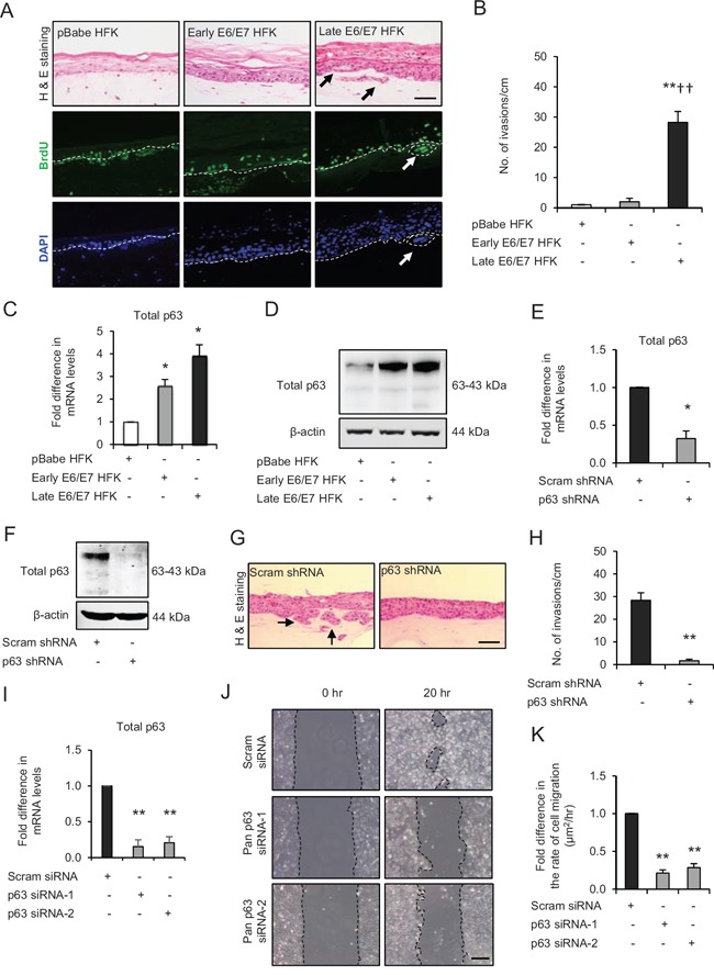
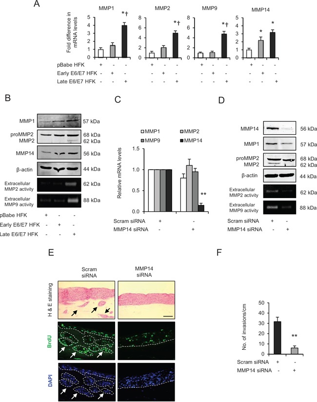
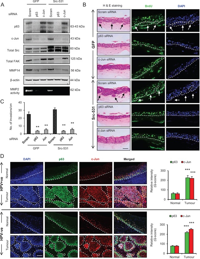
Using microarray information from oro-pharyngeal data sets and results from primary human foreskin keratinocytes (HFK) expressing Human Papilloma Virus (HPV)-16 E6/E7 proteins, we show that p63 expression regulates signalling molecules which initiate cell migration such as Src and focal adhesion kinase (FAK) and induce invasion in 3D-organotypic rafts; a phenotype that can be reversed by depletion of p63. Knockdown of Src or FAK in the invasive cells restored focal adhesion protein paxillin at cell periphery and impaired the cell migration. In addition, specific inhibition of FAK (PF573228) or Src (dasatinib) activities mitigated invasion and attenuated the expression/activity of matrix metalloproteinase 14 (MMP14), a pivotal MMP in the MMP activation cascade. Expression of constitutively active Src in non-invasive HFK expressing E6/E7 proteins upregulated the activity of c-Jun and MMP14, and induced invasion in rafts. Depletion of Src, FAK or AKT in the invasive cells normalised the expression/activity of c-Jun and MMP14, thus implicating the Src-FAK/AKT/AP-1 signalling in MMP14-mediated extra-cellular matrix remodelling. Up-regulation of Src, AP-1, MMP14 and p63 expression was confirmed in oro-pharyngeal cancer. Since p63 transcriptionally regulated expression of many of the genes in this signalling pathway, it suggests that it has a central role in cancer progression.

Keywords: HPV16; MMP14; Src; invasion; p63.


Conflict of interest statement

CONFLICTS OF INTEREST

None

Figures

Figure 1
Figure 1. p63 transcription factors drive cell migration and invasion in late passage human foreskin keratinocytes (HFK) expressing human papilloma virus (HPV)16 E6/E7 genes
A. H&E staining and immunofluorescence detection of BrdU uptake and nuclei (DAPI) on 3D-organotypic rafts established from normal HFK expressing pBabe (control), early passage and late passage HFK expressing E6/E7 genes cells seeded on retinoblastoma-depleted human foreskin fibroblast embedded collagen-I plugs. Invasive incidents are indicated by arrows. B. Quantification of number of invasive incidents across the rafts per cm. C. The relative mRNA and D. protein expression of total p63 and loading control β-actin in aforementioned cells. E. Relative mRNA and F. protein levels of total p63 in late passage E6/E7-HFK after stable p63 (p63 shRNA) knockdown compared to its controls (scram shRNA). G. H&E staining of rafts established from late passage HFK expressing control and stable p63 knockdown. H. Quantification of number of invasive incidents per cm across the rafts. I. Relative mRNA levels of total p63 after transient knockdown by control and two different p63 siRNA molecules in late passage E6/E7-HFK. J. Representative phase contrast images of scratch wound assay, showing cell migration after 20 hr in these cells seeded on collagen-I coated plates. K. Quantification of migration represented as μm2/hr. Scale bars represent 100 μm. N = 3 independent experiments, mean ± SEM, **p < 0.01 compared to the respective controls, *p < 0.05 compared to the control, ††p < 0.01 compared to early passage E6/E7-HFK.
Figure 2
Figure 2. p63 transcription factors modulate Src-focal adhesion kinase (FAK) signalling
A. Microarray data of total p63, Src and PTK2/FAK mRNA levels in normal tissue and HPV-negative (p16-) and HPV-positive (p16+) oro-pharyngeal tumours. B. Correlation analysis of Src and PTK2/FAK against p63 in samples from (A). C. Relative mRNA levels of Src, PTK2/FAK and paxillin in human foreskin keratinocytes (HFK) expressing pBabe (control), early and late passage HFK expressing E6/E7 genes. D. Protein levels of total Src, FAK, paxillin, pSrc-Y416, pFAK-Y397, pFAK-Y576/577 and pPaxillin-Y118; E. cellular localisation (arrows) of total paxillin and pFAK-Y397 in aforementioned cells. F. Relative mRNA levels of PTK2/FAK and Src in p63 depleted cells and G. protein levels of total p63, Src, FAK, paxillin and pPaxillin-Y118 after transient knockdown by Scram (control) and two different p63 siRNA molecules in late passage E6/E7-HFK. β-actin served as loading control. Scale bars represent 50 μm. N = 3 independent experiments, mean ± SEM, ***p < 0.0001 compared to normal, ##p < 0.01 compared to the control, *p < 0.01 compared to the control, ††p < 0.01 compared to early passage E6/E7 HFK, ns: not significant.
Figure 3
Figure 3. Src-focal adhesion kinase (FAK) signalling regulates cell migration and invasion
A. Relative mRNA levels of PTK2/FAK and Src and, B. protein levels of total FAK, Src, paxillin, pSrc-Y416, pFAK-Y397, pPaxillin-Y118 and β-actin after transient knockdown by Scram (control), FAK and Src siRNA molecules in late passage human foreskin keratinocytes (HFK) expressing HPV16 E6/E7 genes. C. Representative phase contrast images of scratch wound assay on collagen-I coated plates showing cell migration after transient knockdown with scram, FAK and Src siRNA in the late passage E6/E7-HFK, which was D. quantified and represented as μm2/hr. E. H&E staining and immunofluorescence detection of BrdU uptake and nuclei (DAPI) on 3D-organotypic rafts established from late passage E6/E7-HFK seeded on retinoblastoma-depleted HFF embedded collagen-I plugs and treated with specific inhibitors of FAK (PF573228) and Src (dasatinib) activities. Rafts treated with DMSO served as control. Invasive incidents are indicated by arrows. F. Quantification of number of invasive incidents across the rafts per cm. Scale bars represents 100 μm. N = 3 independent experiments, mean ± SEM, **p < 0.01 compared to the respective controls, *p < 0.05 compared to the control.
Figure 4
Figure 4. Matrix metalloproteinases (MMP)-mediated extra cellular matrix (ECM) remodelling is important during invasion
A. The relative mRNA levels of MMP1, 2, 9 and 14, B. protein expression and activities on gelatin zymographs of MMPs in human foreskin keratinocytes (HFK) expressing pBabe (control), early and late passage HFK expressing HPV16 E6/E7 genes. C. Relative mRNA levels of MMP1, 2, 9 and 14; and D. protein levels of MMP1, 2, 14 and extracellular activities of MMP2 and MMP9 after transient knockdown by Scram (control) or MMP14 siRNA molecules in the late passage E6/E7-HFK. E. H&E staining and immunofluorescence detection of BrdU uptake and nuclei (DAPI) on 3D-organotypic rafts established from aforementioned MMP14 knockdown cells. Invasive incidents are indicated by arrows. F. Quantification of number of invasive incidents across the rafts per cm. β-actin served as loading control. Scale bar represent 100 μm. N = 3 independent experiments, mean ± SEM, **p < 0.01 compared to the control, *p < 0.05 compared to the controls, p < 0.05 compared to the early passage population.
Figure 5
Figure 5. Src-focal adhesion kinase (FAK) signalling regulates expression and activity of matrix metalloproteinases-14 (MMP14)
A. Relative mRNA levels of MMP14, B. protein levels and activity (observed by extracellular MMP2-mediated digestion of gelatin) of MMP14 after transient knockdown by Scram (control), FAK and Src siRNA molecules in late passage human foreskin keratinocytes (HFK) expressing HPV16 E6/E7 genes. C and D. Protein expression of total MMP14 and MMP14 activity in the late passage populations after dose-dependent inhibition of FAK (PF573228) and Src (dasatinib) activities. E. H&E staining and immunofluorescence detection of BrdU uptake and nuclei (DAPI) on 3D-organotypic rafts established from early passage HFK expressing E6/E7 genes alongside GFP (control), wild type-Src (Src-WT), constitutively active (Src-531) and kinase dead (Src-KD) constructs. The invasive incidents are indicated by arrows. F. Quantification of the number of invasive incidents across the rafts per cm. G. The relative mRNA levels of Src and MMP14 and H. protein levels of V5-tagged Src (Src-531 was not tagged with V5), total Src, pSrc-Y416, total FAK, pFAK-Y576/577, MMP2 and MMP14, and the activity of MMP14 in early passage HFK expressing E6/E7 genes and Src constructs. β-actin served as loading control. Scale bar represent 100 μm. N = 3 independent experiments, mean ± SEM, *p < 0.05 compared to the respective controls, p < 0.05 compared to the early passage population expressing Src-531.
Figure 6
Figure 6. Src-focal adhesion kinase (FAK) signalling regulates the expression and activation of c-Jun via AKT activation
A. The protein levels of pAKT-S473, total AKT, p-c-Jun-S73 and total c-Jun in the normal human foreskin keratinocytes (HFK) expressing pBabe (control), early passage and late passage HFK expressing HPV16 E6/E7 genes. B. Immunofluorescence detection of p-c-Jun-S73 on the rafts. C. The relative mRNA levels of c-Jun and MMP14, and D. protein levels of total AKT, total c-Jun, p-c-Jun-S73, MMP14 and total p63 after transient knockdown with scram (control) and total AKT siRNA in late passage E6/E7-HFK. E. The relative mRNA levels of c-Jun and MMP14, and F. protein levels of total c-Jun, MMP14 and β-actin after transient knockdown with scram (control) and two different c-Jun siRNA molecules in the late passage E6/E7-HFK. G. Protein levels of total FAK, total Src, pAKT-S473, total AKT, p-c-Jun-S73 and total c-Jun after transient knockdown by Scram (control), FAK or Src siRNA molecules in the late passage E6/E7-HFK. H. Immunofluorescence detections of p-c-Jun-S73 and BrdU uptake on 3D-organotypic rafts established from the late passage population and treated with specific inhibitors of FAK (PF573228) and Src (dasatinib) activities. Rafts treated with DMSO served as control. The invasive incidents are indicated by arrows. I. Protein levels of total c-Jun, p-c-Jun-S73 in early passage E6/E7-HFK expressing GFP, wild type-Src (Src-WT), constitutively active (Src-531) and kinase dead (Src-KD) constructs. β-actin served as loading control. Scale bars represent 100 μm. N = 3 independent experiments, mean ± SEM, *p < 0.05 compared to the respective controls, p < 0.05 compared to the respective controls.
Figure 7
Figure 7. p63 regulates the expression and/or activation of AKT, c-Jun and matrix metalloproteinase-14 (MMP14)
A. Protein levels of pAKT-S473, total AKT, total c-Jun, MMP14 and β-actin; and B. the relative mRNA levels of c-Jun and MMP14 after transient knockdown by Scram (control) and two different p63 siRNA molecules in late passage human foreskin keratinocytes (HFK) expressing human papilloma virus (HPV)16 E6/E7 genes. C. Immunofluorescence detections of p-c-Jun-S73 and BrdU uptake on 3D-organotypic rafts established from late passage HFK after stable p63 (p63 shRNA) knockdown compared to its control (scram shRNA). The invasive incidents are indicated by arrows. Scale bar represent 100 μm. N = 3 independent experiments, mean ± SEM, *p < 0.05 compared to the controls.
Figure 8
Figure 8. p63 regulates the Src/c-Jun/matrix metalloproteinases-14 (MMP14)-mediated extra cellular matrix (ECM) remodelling and invasion
A. Protein levels of total p63, c-Jun, Src, focal adhesion kinase (FAK), MMP14 and β-actin; and activity of MMP2 as read out of the MMP14 activity after knockdown with Scram (control), total p63 or c-Jun siRNA in late passage human foreskin keratinocytes (HFK) expressing HPV16 E6/E7 gene alongside GFP or constitutively active Src (Src-531) constructs. B. H&E staining showing invasions (arrows) and immunofluorescence detection of BrdU uptake and nuclei (DAPI) on 3D-organotypic rafts established with cells from (A). C. Quantification of number of invasive incidents across the rafts per cm. D. Immunofluorescence detection and quantification (Q-score) of p63 and c-Jun in normal epithelium and tumour areas from HPV positive (+ve) and HPV negative (−ve) oro-pharyngeal squamous cell carcinomas. Scale bar represent 100 μm. N = 3 independent experiments, 5 (HPV+ve) and 5 (HPV−ve) tumour sections, mean ± SEM, ***p < 0.001 compared to the normal areas, **p < 0.01 compared to the controls.
Figure 9
Figure 9. A. Immunofluorescence detection and quantification (Q-score) of Src and MMP14 in normal epithelium and tumour areas from HPV positive (+ve) and HPV negative (−ve) oro-pharyngeal squamous cell carcinomas
The nuclei are stained with DAPI. B. Schematic representation of the role of p63 proteins in regulating ECM remodelling, cell migration and invasion via Src/FAK/AKT/AP-1 signalling pathway. Scale bar represent 100 μm. N = 5 (HPV+ve) and 5 (HPV−ve) tumour sections, mean ± SEM, ***p < 0.001 compared to the normal areas.

Similar articles

Cited by

References

    1. Ferlay J, Shin H-R, Bray F, Forman D, Mathers C, Parkin DM. Estimates of worldwide burden of cancer in 2008 GLOBOCAN. Int J Cancer. 2008;2010;127:2893–2917. - PubMed
    1. Gillison ML, Koch WM, Capone RB, Spafford M, Westra WH, Wu L, Zahurak ML, Daniel RW, Viglione M, Symer DE, Shah KV, Sidransky D. Evidence for a causal association between human papillomavirus and a subset of head and neck cancers. J Natl Cancer Inst. 2000;92:709–720. - PubMed
    1. McKenna DJ, McDade SS, Patel D, McCance DJ. MicroRNA 203 Expression in Keratinocytes Is Dependent on Regulation of p53 Levels by E6. J Virol. 2010;84:10644–10652. - PMC - PubMed
    1. Melar-New M, Laimins LA. Human Papillomaviruses Modulate Expression of MicroRNA 203 upon Epithelial Differentiation to Control Levels of p63 Proteins. J Virol. 2010;84:5212–5221. - PMC - PubMed
    1. Brunner HG, Hamel BCJ, van Bokhoven H. P63 gene mutations and human developmental syndromes. Am J Med Genet. 2002;112:284–290. - PubMed

MeSH terms

Substances

LinkOut - more resources