

Intraprocedural 3D Quantification of Lipiodol Deposition on Cone-Beam CT Predicts Tumor Response After Transarterial Chemoembolization in Patients with Hepatocellular Carcinoma
- PMID: 26001366
- PMCID: PMC4651808
- DOI: 10.1007/s00270-015-1129-9
Intraprocedural 3D Quantification of Lipiodol Deposition on Cone-Beam CT Predicts Tumor Response After Transarterial Chemoembolization in Patients with Hepatocellular Carcinoma
Abstract
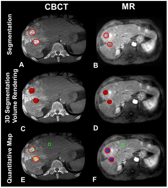
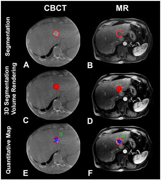
Purpose: To evaluate whether intraprocedural 3D quantification of Lipiodol deposition on cone-beam computed tomography (CBCT) can predict tumor response on follow-up contrast-enhanced magnetic resonance imaging (CE-MRI) in patients with hepatocellular carcinoma (HCC) treated with conventional transarterial chemoembolization (cTACE).
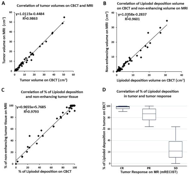
Materials and methods: This IRB approved, retrospective analysis included 36 patients with 51 HCC target lesions, who underwent cTACE with CBCT. CE-MRI was acquired at baseline and 1 month after cTACE. Overall tumor volumes as well as intratumoral Lipiodol volumes on CBCT were measured and compared with the overall and necrotic (non-enhancing) tumor volumes on CE-MRI using the paired student's t test. Tumor response on CE-MRI was assessed using modified response evaluation criteria in solid tumors (mRECIST). A linear regression model was used to correlate tumor volumes, Lipiodol volumes, and the percentage of Lipiodol deposition on CBCT with the corresponding parameters on CE-MRI. Nonparametric spearman rank-order correlation and trend test were used to correlate the percentage of Lipiodol deposition in the tumor with tumor response.
Result: A strong correlation between overall tumor volumes on CBCT and CE-MRI was observed (R(2) = 0.986). In addition, a strong correlation was obtained between the volume of Lipiodol deposition on CBCT and tumor necrosis (in cm(3)) on CE-MRI (R(2) = 0.960), and between the percentage of Lipiodol deposition and tumor necrosis (R(2) = 0.979). Importantly, the extent of Lipiodol deposition (in percentage of total tumor volume) correlated strongly with tumor response on CE-MRI (Spearman rho = 0.84, p < 0.001).
Conclusions: Intraprocedural 3D quantification of Lipiodol deposition on CBCT can be used to predict tumor response on follow-up CE-MRI.
Keywords: Cone-beam computed tomography; Hepatocellular carcinoma; Quantitative; Three dimensional; Transcatheter arterial chemoembolization; Tumor response.
Figures
Similar articles
-
Lipiodol retention pattern assessed by cone beam computed tomography during conventional transarterial chemoembolization of hepatocellular carcinoma: accuracy and correlation with response.Cancer Imaging. 2016 Oct 3;16(1):32. doi: 10.1186/s40644-016-0090-4. Cancer Imaging. 2016. PMID: 27716376 Free PMC article.
-
Quantitative assessment of lipiodol deposition after chemoembolization: comparison between cone-beam CT and multidetector CT.J Vasc Interv Radiol. 2013 Dec;24(12):1837-44. doi: 10.1016/j.jvir.2013.08.017. Epub 2013 Oct 1. J Vasc Interv Radiol. 2013. PMID: 24094672 Free PMC article.
-
Early prediction of 1-year tumor response of hepatocellular carcinoma with lipiodol deposition pattern through post-embolization cone-beam computed tomography during conventional transarterial chemoembolization.Eur Radiol. 2021 Oct;31(10):7464-7475. doi: 10.1007/s00330-021-07843-8. Epub 2021 Mar 25. Eur Radiol. 2021. PMID: 33765160
-
Initiative on Superselective Conventional Transarterial Chemoembolization Results (INSPIRE).Cardiovasc Intervent Radiol. 2022 Oct;45(10):1430-1440. doi: 10.1007/s00270-022-03233-9. Epub 2022 Aug 17. Cardiovasc Intervent Radiol. 2022. PMID: 35978174 Free PMC article. Review.
-
A Review Of Mri Appearances Of Lipiodol In Conventional Tace (Ctace) Treated Hepatocellular Carcinomas.J Ayub Med Coll Abbottabad. 2023 Oct-Dec;35(Suppl 1)(4):S740-S745. doi: 10.55519/JAMC-S4-11966. J Ayub Med Coll Abbottabad. 2023. PMID: 38406903 Review.
Cited by
-
Utility of 'dual phase' cone beam computed tomography during radioembolisation in patients with hepatocellular carcinoma: what is really changing in flow dynamics before and after 90Y delivery?Pol J Radiol. 2020 Jan 14;85:e21-e28. doi: 10.5114/pjr.2020.92915. eCollection 2020. Pol J Radiol. 2020. PMID: 32180850 Free PMC article.
-
Single injection dual phase CBCT technique ameliorates results of trans-arterial chemoembolization for hepatocellular cancer.Transl Gastroenterol Hepatol. 2017 Oct 24;2:83. doi: 10.21037/tgh.2017.10.03. eCollection 2017. Transl Gastroenterol Hepatol. 2017. PMID: 29167830 Free PMC article. Review.
-
Bench-to-clinic development of imageable drug-eluting embolization beads: finding the balance.Future Oncol. 2018 Nov;14(26):2741-2760. doi: 10.2217/fon-2018-0196. Epub 2018 Jun 26. Future Oncol. 2018. PMID: 29944007 Free PMC article. Review.
-
Theranostics in Interventional Oncology: Versatile Carriers for Diagnosis and Targeted Image-Guided Minimally Invasive Procedures.Front Pharmacol. 2019 May 9;10:450. doi: 10.3389/fphar.2019.00450. eCollection 2019. Front Pharmacol. 2019. PMID: 31143114 Free PMC article. Review.
-
Arms Down Cone Beam CT Hepatic Angiography Performance Assessment: Vascular Imaging Quality and Imaging Artifacts.Cardiovasc Intervent Radiol. 2018 Jun;41(6):898-904. doi: 10.1007/s00270-017-1875-y. Epub 2018 Jan 11. Cardiovasc Intervent Radiol. 2018. PMID: 29327076 Free PMC article.
References
-
- Llovet JM, Real MI, Montana X, Planas R, Coll S, Aponte J, et al. Arterial embolisation or chemoembolisation versus symptomatic treatment in patients with unresectable hepatocellular carcinoma: a randomised controlled trial. Lancet. 2002;359(9319):1734–9. doi: 10.1016/S0140-6736(02)08649-X. PMID: 12049862. - PubMed
-
- Lo CM, Ngan H, Tso WK, Liu CL, Lam CM, Poon RT, et al. Randomized controlled trial of transarterial lipiodol chemoembolization for unresectable hepatocellular carcinoma. Hepatology. 2002;35(5):1164–71. doi: 10.1053/jhep.2002.33156. PMID: 11981766. - PubMed
-
- Herber S, Biesterfeld S, Franz U, Schneider J, Thies J, Schuchmann M, et al. Correlation of multislice CT and histomorphology in HCC following TACE: predictors of outcome. Cardiovascular and interventional radiology. 2008;31(4):768–77. doi: 10.1007/s00270-007-9270-8. PMID: 18196335. - PubMed
-
- Takayasu K, Muramatsu Y, Maeda T, Iwata R, Furukawa H, Muramatsu Y, et al. Targeted transarterial oily chemoembolization for small foci of hepatocellular carcinoma using a unified helical CT and angiography system: analysis of factors affecting local recurrence and survival rates. AJR American journal of roentgenology. 2001;176(3):681–8. doi: 10.2214/ajr.176.3.1760681. PMID: 11222205. - PubMed
-
- Loffroy R, Lin M, Yenokyan G, Rao PP, Bhagat N, Noordhoek N, et al. Intraprocedural C-arm dual-phase cone-beam CT: can it be used to predict short-term response to TACE with drug-eluting beads in patients with hepatocellular carcinoma? Radiology. 2013;266(2):636–48. doi: 10.1148/radiol.12112316. PMID: 23143027. - PMC - PubMed
MeSH terms
Substances
Grants and funding
LinkOut - more resources
Full Text Sources
Other Literature Sources
Medical
