Characterization of single-domain antibodies against Foot and Mouth Disease Virus (FMDV) serotype O from a camelid and imaging of FMDV in baby hamster kidney-21 cells with single-domain antibody-quantum dots probes
- PMID: 26001568
- PMCID: PMC4489003
- DOI: 10.1186/s12917-015-0437-2
Characterization of single-domain antibodies against Foot and Mouth Disease Virus (FMDV) serotype O from a camelid and imaging of FMDV in baby hamster kidney-21 cells with single-domain antibody-quantum dots probes
Abstract
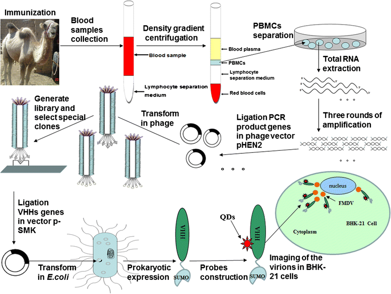
Background: Foot-and-mouth disease (FMD) is a highly contagious disease that affects cloven-hoofed animals and causes significant economic losses to husbandry worldwide. The variable domain of heavy-chain antibodies (VHHs or single domain antibodies, sdAbs) are single-domain antigen-binding fragments derived from camelid heavy-chain antibodies.
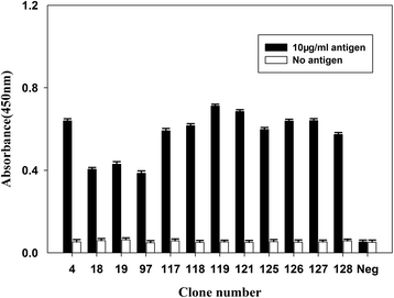
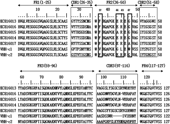
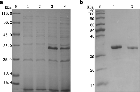
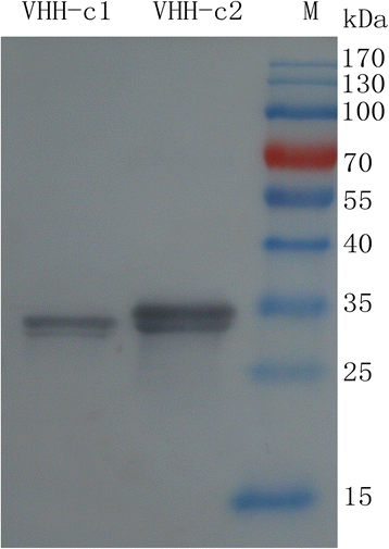
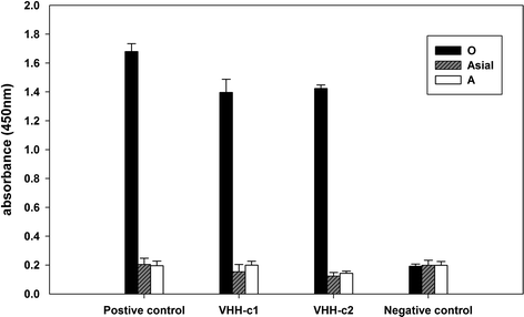
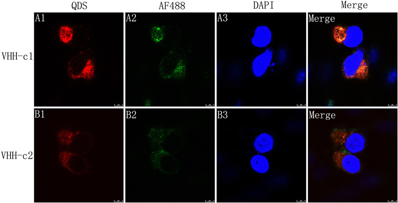
Results: In this work, two sdAbs against FMD virus (FMDV) serotype O were selected from a camelid phage display immune library and expressed in Escherichia coli. The serotype specificity and affinity of the sdAbs were identified through enzyme-linked immunosorbent assay and surface plasmon resonance assay. Moreover, the sdAbs were conjugated with quantum dots to constitute probes for imaging FMD virions. Results demonstrated that the two sdAbs were specific for serotype O and shared no cross-reactivity with serotypes A and Asia 1. The equilibrium dissociation constant (KD) values of the two sdAbs ranged from 6.23 nM to 8.24 nM, which indicated high affinity to FMDV antigens. Co-localization with the sdAb-AF488 and sdAb-QD probes indicated the same location of FMDV virions in baby hamster kidney-21 (BHK-21) cells.
Conclusions: sdAb-QD probes are powerful tools to detect and image FMDV in BHK-21 cells.
Figures
Similar articles
-
Characterization of Asia 1 sdAb from camels bactrianus (C. bactrianus) and conjugation with quantum dots for imaging FMDV in BHK-21 cells.PLoS One. 2013 May 30;8(5):e63500. doi: 10.1371/journal.pone.0063500. Print 2013. PLoS One. 2013. PMID: 23737944 Free PMC article.
-
Passive immunization of guinea pigs with llama single-domain antibody fragments against foot-and-mouth disease.Vet Microbiol. 2007 Mar 10;120(3-4):193-206. doi: 10.1016/j.vetmic.2006.10.029. Epub 2006 Oct 28. Vet Microbiol. 2007. PMID: 17127019
-
Serotype-independent detection of foot-and-mouth disease virus.J Virol Methods. 2008 Jul;151(1):146-53. doi: 10.1016/j.jviromet.2008.03.011. Epub 2008 Apr 25. J Virol Methods. 2008. PMID: 18440078
-
Development of vaccines toward the global control and eradication of foot-and-mouth disease.Expert Rev Vaccines. 2011 Mar;10(3):377-87. doi: 10.1586/erv.11.4. Expert Rev Vaccines. 2011. PMID: 21434805 Review.
-
Enhancing Stability of Camelid and Shark Single Domain Antibodies: An Overview.Front Immunol. 2017 Jul 25;8:865. doi: 10.3389/fimmu.2017.00865. eCollection 2017. Front Immunol. 2017. PMID: 28791022 Free PMC article. Review.
Cited by
-
Single-domain antibodies as promising experimental tools in imaging and isolation of porcine epidemic diarrhea virus.Appl Microbiol Biotechnol. 2018 Oct;102(20):8931-8942. doi: 10.1007/s00253-018-9324-7. Epub 2018 Aug 24. Appl Microbiol Biotechnol. 2018. PMID: 30143837 Free PMC article.
-
Selection and identification of single-domain antibody against Peste des Petits Ruminants virus.J Vet Sci. 2021 Jul;22(4):e45. doi: 10.4142/jvs.2021.22.e45. Epub 2021 Jun 2. J Vet Sci. 2021. PMID: 34170088 Free PMC article.
-
Identification and characterization of nanobodies specifically against African swine fever virus major capsid protein p72.Front Microbiol. 2022 Oct 13;13:1017792. doi: 10.3389/fmicb.2022.1017792. eCollection 2022. Front Microbiol. 2022. PMID: 36312984 Free PMC article.
-
Developing Recombinant Antibodies by Phage Display Against Infectious Diseases and Toxins for Diagnostics and Therapy.Front Cell Infect Microbiol. 2021 Jul 7;11:697876. doi: 10.3389/fcimb.2021.697876. eCollection 2021. Front Cell Infect Microbiol. 2021. PMID: 34307196 Free PMC article. Review.
-
Antibody Phage Display Technology for Sensor-Based Virus Detection: Current Status and Future Prospects.Biosensors (Basel). 2023 Jun 9;13(6):640. doi: 10.3390/bios13060640. Biosensors (Basel). 2023. PMID: 37367005 Free PMC article. Review.
References
-
- Yang PC, Chu RM, Chung WB, Sung HT. Epidemiological characteristics and financial costs of the 1997 foot-and-mouth disease epidemic in Taiwan. Vet Rec. 1999;145:731–4. - PubMed
-
- Thompson D, Muriel P, Russell D, Osborne P, Bromley A, Rowland M, et al. Economic costs of the foot and mouth disease outbreak in the United Kingdom in 2001. Rev Sci Tech. 2002;21:675–87. - PubMed
-
- Garland A, Donaldson A. Foot-and-mouth disease. Surveillance. 1990;17:6–8.
Publication types
MeSH terms
Substances
LinkOut - more resources
Full Text Sources
Other Literature Sources

