Review
. 2015 Sep;72(17):3267-80.
doi: 10.1007/s00018-015-1926-y. Epub 2015 May 23.

Cytoplasmic dynein and early endosome transport

Affiliations
Review

Cytoplasmic dynein and early endosome transport

Xin Xiang et al. Cell Mol Life Sci. 2015 Sep.

Abstract

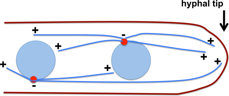
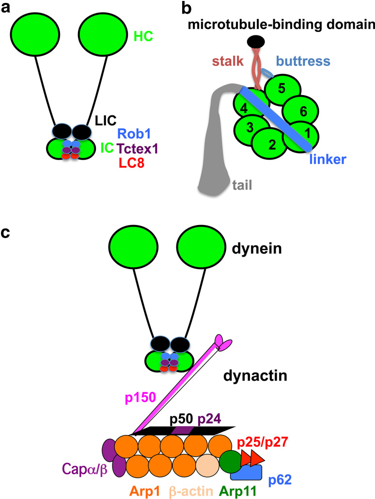
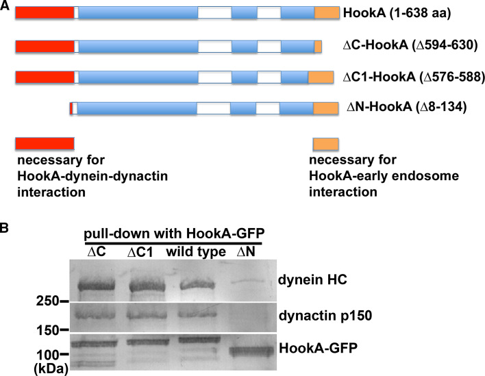
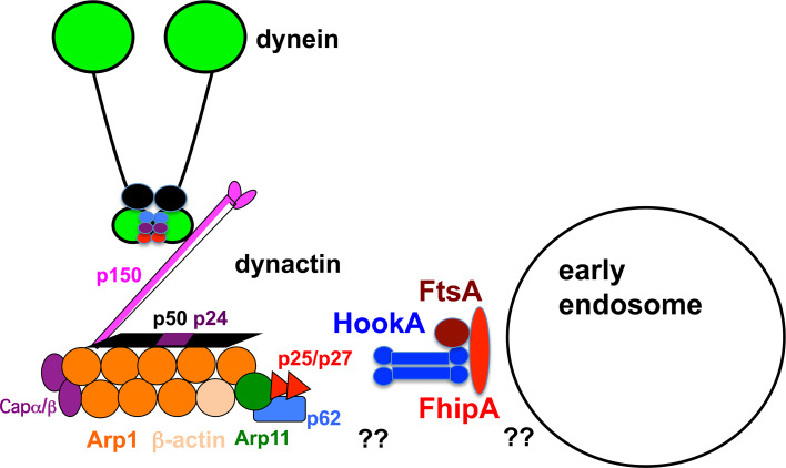
Microtubule-based distribution of organelles/vesicles is crucial for the function of many types of eukaryotic cells and the molecular motor cytoplasmic dynein is required for transporting a variety of cellular cargos toward the microtubule minus ends. Early endosomes represent a major cargo of dynein in filamentous fungi, and dynein regulators such as LIS1 and the dynactin complex are both required for early endosome movement. In fungal hyphae, kinesin-3 and dynein drive bi-directional movements of early endosomes. Dynein accumulates at microtubule plus ends; this accumulation depends on kinesin-1 and dynactin, and it is important for early endosome movements towards the microtubule minus ends. The physical interaction between dynein and early endosome requires the dynactin complex, and in particular, its p25 component. The FTS-Hook-FHIP (FHF) complex links dynein-dynactin to early endosomes, and within the FHF complex, Hook interacts with dynein-dynactin, and Hook-early endosome interaction depends on FHIP and FTS.


Figures

Fig. 1
Fig. 1
A schematic diagram showing microtubule organization in multinucleated fungi such as A. nidulans. Blue circles nuclei. Blue lines microtubules. Red circles spindle-pole bodies. A microtubule plus end is labeled as “+” and minus end as “−”. In the middle of hyphae, microtubules are of mixed polarity, but in a region close to the hyphal tip, the microtubule plus ends face the hyphal apex
Fig. 2
Fig. 2
Schematic diagrams of the cytoplasmic dynein complex and the dynactin complex. a The dynein complex. This diagram was modified from [60]. The dynein heavy chain motor (HC) and other subunits, including the intermediate chain (IC), light intermediate chain (LIC) and three families of light chains (Rob1, Tctex1, LC8) are shown. b Different domains of the dynein heavy chain. This diagram was modified from [67]. Each heavy chain (HC) contains the N-terminal tail and the C-terminal motor unit with six AAA domains (marked 1–6) that are organized in a ring-like structure. The linker is an important mechanical element connected to AAA1. The coiled-coil stalk extends out from AAA4 and leads to the microtubule-binding domain. The buttress is a coiled-coil hairpin that extends out of AAA5 and contacts the stalk. c A simplified diagram of dynein–dynactin when the two complexes are together. The dynactin complex is modified from a previous publication [11]. The interaction between the dynein IC and p150 dynactin is depicted [–95], but the recently identified interaction between the HC tail and Arp1 is not depicted, and more detailed information on dynactin structure can be found in the recent publications [88, 96]
Fig. 3
Fig. 3
Hook-dynein-dynactin interaction requires the N-terminal region but not the C-terminal cargo-binding domain of Hook. Here we present data obtained from A. nidulans Hook (HookA) to show this point. a A diagram of HookA, ∆C-HookA, ∆C1-HookA and ∆N-HookA. Red box N-terminal region, Blue boxes coiled-coil domains, Brown box the C-terminal domain. The functions of the N- and C-terminus are indicated at the bottom. Note that in the ∆C1 mutant, 13 aa at the end of the third coiled-coil are deleted, which disrupts the function of the C-terminal domain in early endosome binding. b The dynein HC and the p150 subunit of dynactin were pulled down with HookA-GFP, which requires the N-terminus but not the C-terminus of HookA. This figure was modified from [15]
Fig. 4
Fig. 4
Model showing the FTS–Hook–FHIP complex (FtsA-HookA-FhipA in A. nidulans) linking dynein–dynactin to early endosomes. HookA (blue, depicted as a dimer [175] interacts with dynein–dynactin complexes [13, 15], but the mechanism of the interaction is not clear (indicated by double question marks). The C-terminus of HookA interacts with FtsA (brown) [175]. FhipA (red) is depicted to be most close to early endosome [14]. Missing linkers are indicated by double question marks

Similar articles

Cited by

References

    1. Engqvist-Goldstein AE, Drubin DG. Actin assembly and endocytosis: from yeast to mammals. Annu Rev Cell Dev Biol. 2003;19:287–332. - PubMed
    1. Maldonado-Baez L, Williamson C, Donaldson JG. Clathrin-independent endocytosis: a cargo-centric view. Exp Cell Res. 2013;319:2759–2769. - PMC - PubMed
    1. Mooren OL, Galletta BJ, Cooper JA. Roles for actin assembly in endocytosis. Annu Rev Biochem. 2012;81:661–686. - PubMed
    1. Schmid SL, Sorkin A, Zerial M. Endocytosis: past, present, and future. Cold Spring Harb Perspect Biol. 2014;6:a022509. - PMC - PubMed
    1. Peñalva MA. Endocytosis in filamentous fungi: Cinderella gets her reward. Curr Opin Microbiol. 2010;13:684–692. - PubMed

Publication types