. 2015 Aug 1;75(15):3167-80.
doi: 10.1158/0008-5472.CAN-14-3701. Epub 2015 May 22.

TP53 Silencing Bypasses Growth Arrest of BRAFV600E-Induced Lung Tumor Cells in a Two-Switch Model of Lung Tumorigenesis

Affiliations

TP53 Silencing Bypasses Growth Arrest of BRAFV600E-Induced Lung Tumor Cells in a Two-Switch Model of Lung Tumorigenesis

Anny Shai et al. Cancer Res. .

Abstract

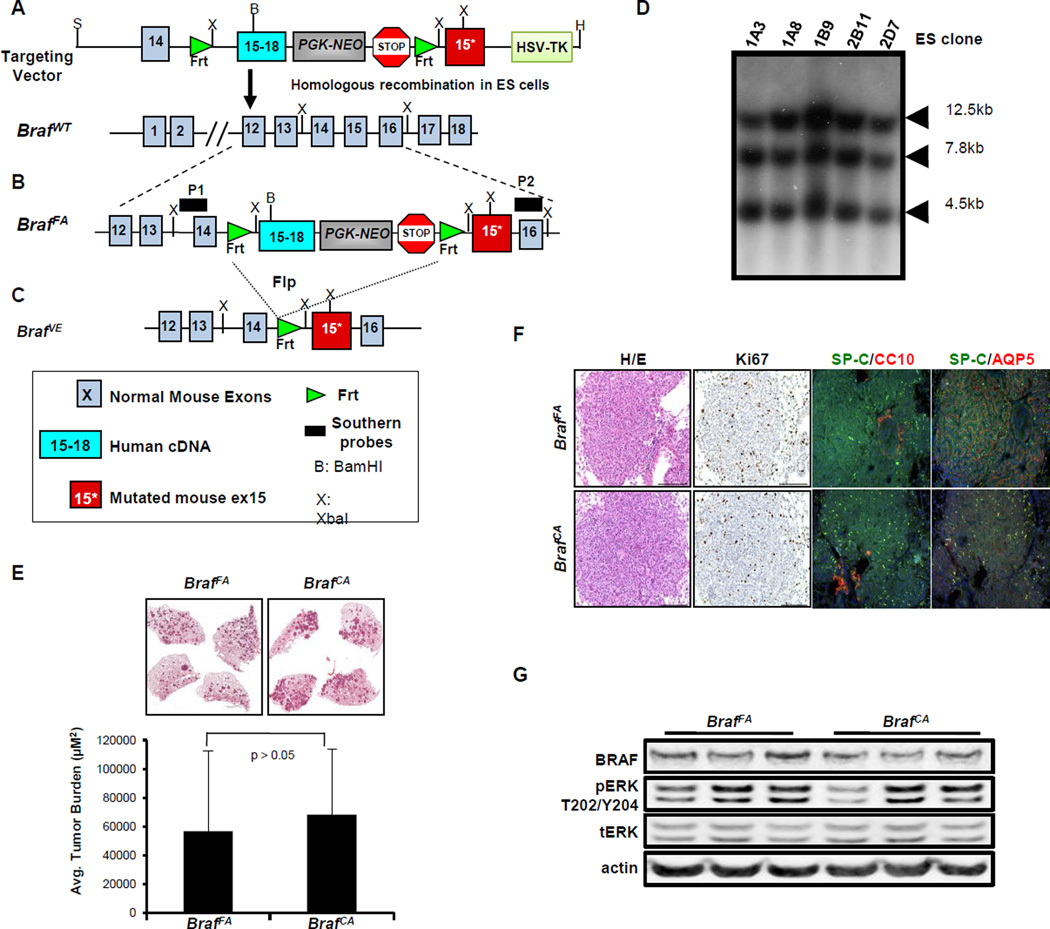
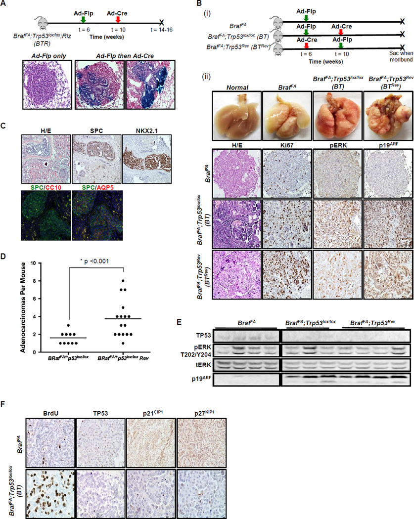
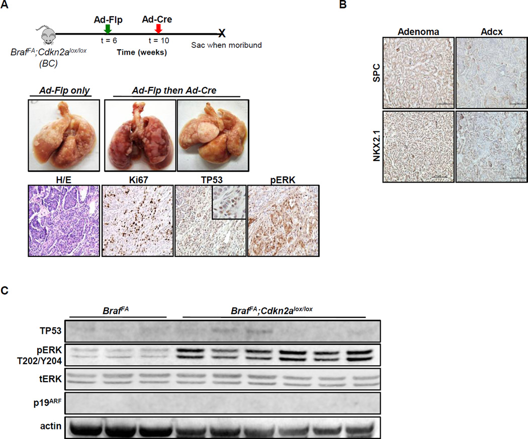
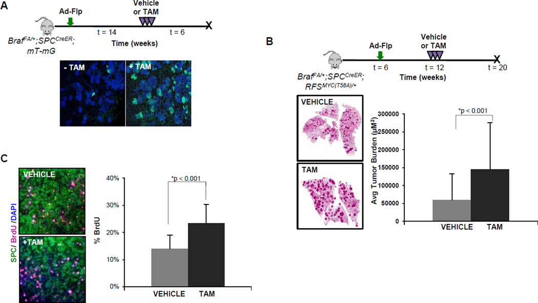
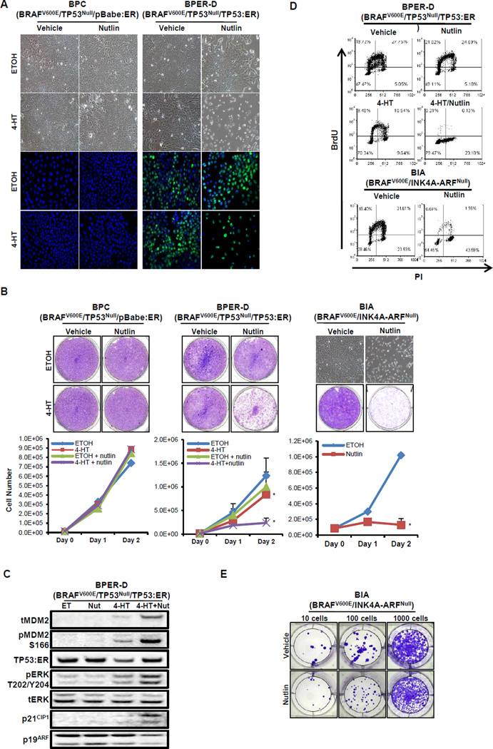
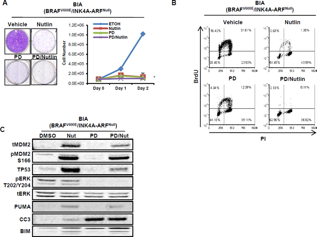
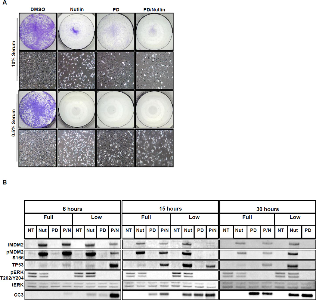
Lung carcinogenesis is a multistep process in which normal lung epithelial cells are converted to cancer cells through the sequential acquisition of multiple genetic or epigenetic events. Despite the utility of current genetically engineered mouse (GEM) models of lung cancer, most do not allow temporal dissociation of the cardinal events involved in lung tumor initiation and cancer progression. Here we describe a novel two-switch GEM model for BRAF(V600E)-induced lung carcinogenesis allowing temporal dissociation of these processes. In mice carrying a Flp recombinase-activated allele of Braf (Braf(FA)) in conjunction with Cre-regulated alleles of Trp53, Cdkn2a, or c-MYC, we demonstrate that secondary genetic events can promote bypass of the senescence-like proliferative arrest displayed by BRAF(V600E)-induced lung adenomas, leading to malignant progression. Moreover, restoring or activating TP53 in cultured BRAF(V600E)/TP53(Null) or BRAF(V600E)/INK4A-ARF(Null) lung cancer cells triggered a G1 cell-cycle arrest regardless of p19(ARF) status. Perhaps surprisingly, neither senescence nor apoptosis was observed upon TP53 restoration. Our results establish a central function for the TP53 pathway in restricting lung cancer development, highlighting the mechanisms that limit malignant progression of BRAF(V600E)-initiated tumors.


Figures

Figure 1
Figure 1. Generation of BrafFA mice
A. Schematic representation of the targeting construct used to generate the BrafFA mouse through homologous recombination into the endogenous Braf genomic locus of 129SvEv derived ES cells. B. Schematic of the structure of the BrafFA allele that expresses normal BRAF and NEO prior to Flp-mediated recombination. P1 and P2 indicate the location of Southern blotting probes used to confirm appropriate targeting of the Braf gene. C. Schematic of the structure of the BrafFA allele following Flp-mediated recombination that now expresses mutationally activated BRAFV600E. D. Southern blot analysis to identify ES clones with the BrafFA targeting construct. E. H&E stained sections of BrafFA (left) and BrafCA (right) 10 weeks p.i. with Ad-Flp or Ad-Cre respectively with average tumor burden indicated in the bar graph. F. Lung sections of tumor-bearing BrafFA or BrafCA mice stained with H&E, Ki67, anti-SPC/CC10 or AQP5 using either IHC and IF methods as indicated. G. Immunoblot analysis of lung tumor lysates from BrafFA or BrafCA mice 10 weeks p.i.
Figure 2
Figure 2. Temporal dissociation of BRAFV600E expression from additional genetic events
A. Lung infection schematic of BrafFA;Trp53lox/lox; Rlz (BTR) mice to test the efficiency of Ad-Cre mediated recombination in established BRAFV600E-driven tumors initiated with Ad-Flp. Below are H&Es stained with β-galactosidase. B. (i) Schematic of infection (ii) Comparison of temporal separation of TP53 silencing and BRAFV600E activation to BRAFV600E alone using two infection strategies. 1st row, photographs of the macro lung. 2nd–4th rows, paraffin lung sections of BrafFA, BT and BrafFA; Trp53Rev (BTRev) stained with H&E, Ki67, pERK, and p19ARF to compare proliferation and MAPK signaling. C. Paraffin sections of a kidney metastasis detected in a BTRev mouse stained with H&E, SPC, NKX2.1, CC10 and AQP5 by IHC and IF. D. Dot plot showing the average number of lung adenocarcinomas in BT or BTRev mice in which BRAFV600E was activated either prior to or after silencing of TP53. E. Immunoblot analysis of BRAFV600E or BRAFV600E/TP53Null lung tumor lysates as indicated. F. Sections of lung tumors from BrafFA or BT mice were stained to detect BrdU incorporation or for expression of TP53, p21CIP1 or p27KIP1 as indicated.
Figure 3
Figure 3. Temporal dissociation of INK4A-ARF silencing cooperates with BRAFV600E to promote lung carcinogenesis
A. (Upper panels) Photographs of the macroscopic lungs from mice of the indicated genotypes. (Lower panels) FFPE lung sections from BrafFA;Cdkn2aflox/flox (BC) mice stained with H&E, or anti-Ki67, -TP53 or pERK by IHC. B. Lung adenomas and adenocarcinomas from BC mice stained with antisera to detect expression of SPC or NKX2.1. C. Immunoblot analysis of lung tumor lysates from BrafFA or BC mice as indicated.
Figure 4
Figure 4. High efficiency Cre-mediated recombination in Ad-Flp initiated BRAFV600E-driven tumors using a SPCCreER transgene
A. Lung tumorigenesis was initiated in BrafFA;SPCCreER;mT-mG mice using Ad-Flp. 14 weeks p.i. mice were either untreated (-TAM) or treated with tamoxifen (+TAM). Six weeks later, mice were euthanized with Cre-mediated expression EGFP in lung tumors assessed by IF. B. Lung tumorigenesis was initiated in 6 week old BrafFA;SPCCreER;RFSMYC(T58A) mice with Ad-Flp. Mice were either treated with vehicle or with tamoxifen (+TAM) 6 weeks p.i. and euthanized with lung tumor burden assessed. C. Sections of tumor bearing lungs from BrafFA;SPCCreER;RFSMYC(T58A) mice treated with vehicle or TAM and administered BrdU prior to sacrifice. Sections were stained as indicated and BrdU positive cells were quantified below.
Figure 5
Figure 5. Restoration of TP53 function in BRAFV600E/TP53Null lung tumor cells results in a G1 cell cycle arrest
A. Phase contrast photomicrographs of BRAFV600E/TP53Null cells infected with either a control (BPC) or a TP53:ER (BPER-D) encoding pBabePuro retrovirus vector. Cells were then treated with 4-hydroxytamoxifen (4-HT) to activate TP53:ER in the absence or presence of Nutlin-3a for 48 hours as indicated. The expression of TP53:ER (green) was assessed by IF. B. BPC or BPER-D cells were treated with vehicle or 4-HT in the absence or presence of nutlin-3a as indicated with cell proliferation assessed using Crystal Violet staining or by cell counting (lower panels). BRAFV600E/INK4A-ARFNull (BIA) lung tumor cells were treated with vehicle or nutlin-3a as indicated with cell proliferation assessed using Crystal Violet staining or by cell counting. A 2-sided t-test was performed to assess significance (* and ** indicates p < 0.05 and p<0.001 respectively). C. Immunoblot analysis of BPER-D cell lysates treated with ethanol (ET) as vehicle control, nutlin-3a (Nut), 4-HT or the combination as indicated. D. Analysis of BrdU incorporation by flow cytometry in BPER-D or BIA cells after 48 hours of treatment with the indicated agents. E. Crystal violet staining of BIA cells replated and cultured for 5 days following treatment with either vehicle or nutlin-3a for 48 hours.
Figure 6
Figure 6. Inhibition of BRAFV600E/INK4A-ARFNull lung cancer cell growth by either MEK or MDM2 inhibition
A. BRAFV600E/INK4A-ARFNull (BIA) lung cancer cells were treated with vehicle, nutlin-3a, MEK1/2 inhibitor (PD325901, PD) or the combination with cell proliferation assessed by staining with Crystal Violet or cell counting. A 2-sided t-test was performed to assess significance (* indicates p <0.001). All three conditions are significant versus vehicle, as well as combination treatment vs single agent. B. BIA lung cancer cells were treated with vehicle, nutlin-3a, MEK1/2 inhibitor (PD) or the combination with transit through S phase of the cell division cycle assessed by labeling with BrdU. BrdU incorporation and position in the cell cycle after 24 hours of drug treatment was assessed by co-staining with an anti-BrdU antisera and propidium iodide and then detected by iF and flow cytometry. C. Immunoblot analysis of BIA lung cancer cells treated with vehicle, nutlin-3a, MEK1/2 inhibitor (PD) or the combination as indicated.
Figure 7
Figure 7. Serum growth factors influence the response of cells to inhibition of MDM2
A. BIA lung cancer cells were treated with vehicle, nutlin-3a, MEK1/2 inhibitor (PD) or the combination in either 0.5%(v/v) or 10%(v/v) fetal calf serum with viable cells stained with Crystal Violet. Photomicrographs of cells in each treatment condition are also provided. B. Immunoblot analysis of BIA cells grown in either high (10%) or low (0.5%) fetal calf serum and then treated with either control, nutlin, PD or the combination for 6, 15 or 30 hours. (NB: No sample could be harvested from BIA cells treated with the combination of nutlin-3a plus PD for 30 hours in low serum).

Similar articles

Cited by

References

    1. Hayat MJ, Howlader N, Reichman ME, Edwards BK. Cancer statistics, trends, and multiple primary cancer analyses from the Surveillance, Epidemiology, and End Results (SEER) Program. Oncologist. 2007;12(1):20–37. - PubMed
    1. Imielinski M, Greulich H, Kaplan B, Araujo L, Amann J, Horn L, et al. Oncogenic and sorafenib-sensitive ARAF mutations in lung adenocarcinoma. J Clin Invest. 2014;124(4):1582–1586. - PMC - PubMed
    1. Heist RS, Engelman JA. SnapShot: non-small cell lung cancer. Cancer Cell. 2012;21(3):448 e2. - PubMed
    1. Network CGAR. Comprehensive molecular profiling of lung adenocarcinoma. Nature. 2014;511(7511):543–550. - PMC - PubMed
    1. Cerami E, Gao J, Dogrusoz U, Gross BE, Sumer SO, Aksoy BA, et al. The cBio cancer genomics portal: an open platform for exploring multidimensional cancer genomics data. Cancer Discov. 2012;2(5):401–404. - PMC - PubMed

Publication types

MeSH terms