

An approach for determining quantitative measures for bone volume and bone mass in the pediatric spina bifida population
- PMID: 26002057
- PMCID: PMC4523422
- DOI: 10.1016/j.clinbiomech.2015.04.010
An approach for determining quantitative measures for bone volume and bone mass in the pediatric spina bifida population
Abstract
Background: The pediatric spina bifida population suffers from decreased mobility and recurrent fractures. This study aimed to develop a method for quantifying bone mass along the entire tibia in youth with spina bifida. This will provide information about all potential sites of bone deficiencies.
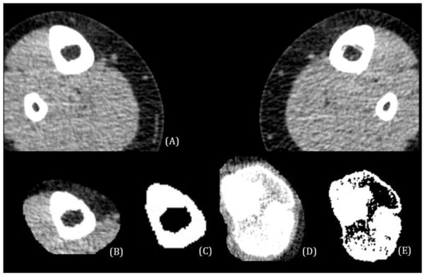
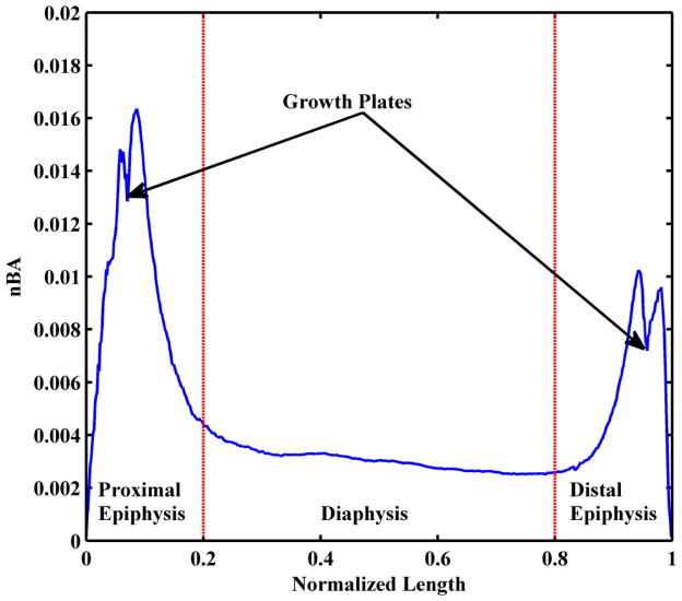
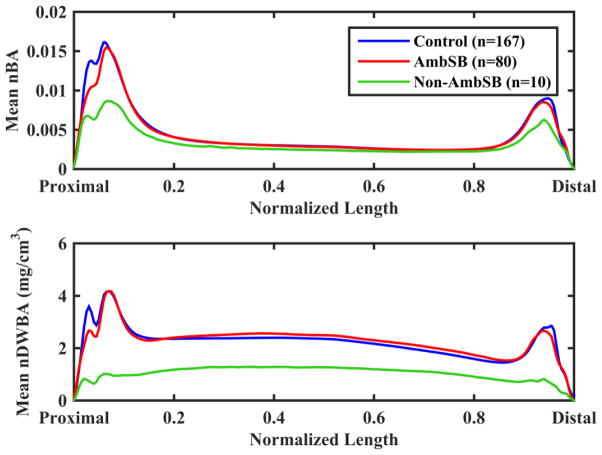
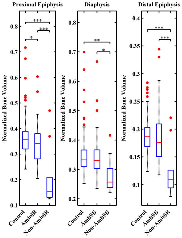
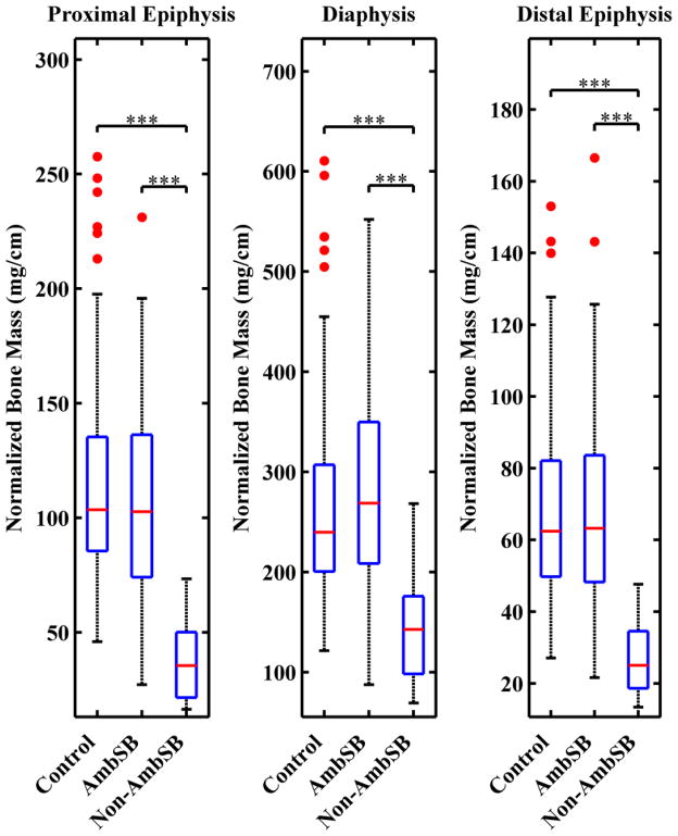
Methods: Computed tomography images of the tibia for 257 children (n=80 ambulatory spina bifida, n=10 non-ambulatory spina bifida, n=167 typically developing) were analyzed. Bone area was calculated at regular intervals along the entire tibia length and then weighted by calibrated pixel intensity for density weighted bone area. Integrals of density weighted bone area were used to quantify bone mass in the proximal and distal epiphyses and diaphysis. Group differences were evaluated using analysis of variance.
Findings: Non-ambulatory children suffer from decreased bone mass in the diaphysis and proximal and distal epiphyses compared to ambulatory and control children (P≤0.001). Ambulatory children with spina bifida showed statistically insignificant differences in bone mass in comparison to typically developing children at these sites (P>0.5).
Interpretation: This method provides insight into tibial bone mass distribution in the pediatric spina bifida population by incorporating information along the whole length of the bone, thereby providing more information than dual-energy x-ray absorptiometry and peripheral quantitative computed tomography. This method can be applied to any population to assess bone mass distribution across the length of any long bone.
Keywords: Bone density; Bone mass; Image analysis; Myelomeningocele; Spina bifida.
Copyright © 2015 Elsevier Ltd. All rights reserved.
Figures
Similar articles
-
Children with spina bifida are at risk for low bone density.Clin Orthop Relat Res. 2011 May;469(5):1253-7. doi: 10.1007/s11999-010-1634-8. Clin Orthop Relat Res. 2011. PMID: 21042897 Free PMC article.
-
Interpretation of whole body dual energy X-ray absorptiometry measures in children: comparison with peripheral quantitative computed tomography.Bone. 2004 Jun;34(6):1044-52. doi: 10.1016/j.bone.2003.12.003. Bone. 2004. PMID: 15193552
-
Risk of fracture prevention in spina bifida patients: correlation between bone mineral density, vitamin D, and electrolyte values.Childs Nerv Syst. 2015 Aug;31(8):1361-5. doi: 10.1007/s00381-015-2726-2. Epub 2015 May 1. Childs Nerv Syst. 2015. PMID: 25930725
-
Bone loss at the distal femur and proximal tibia in persons with spinal cord injury: imaging approaches, risk of fracture, and potential treatment options.Osteoporos Int. 2017 Mar;28(3):747-765. doi: 10.1007/s00198-016-3798-x. Epub 2016 Dec 5. Osteoporos Int. 2017. PMID: 27921146 Review.
-
Update on Urological Management of Spina Bifida from Prenatal Diagnosis to Adulthood.J Urol. 2015 Aug;194(2):288-96. doi: 10.1016/j.juro.2015.03.107. Epub 2015 Apr 1. J Urol. 2015. PMID: 25839383 Review.
Cited by
-
Independently ambulatory children with spina bifida experience near-typical knee and ankle joint moments and forces during walking.Gait Posture. 2023 Jan;99:1-8. doi: 10.1016/j.gaitpost.2022.10.010. Epub 2022 Oct 18. Gait Posture. 2023. PMID: 36283301 Free PMC article.
-
Children with myelomeningocele do not exhibit normal remodeling of tibia roundness with physical development.Bone. 2018 Sep;114:292-297. doi: 10.1016/j.bone.2018.07.001. Epub 2018 Jul 4. Bone. 2018. PMID: 29991457 Free PMC article.
-
Muscle-Bone Interactions in Pediatric Bone Diseases.Curr Osteoporos Rep. 2017 Oct;15(5):425-432. doi: 10.1007/s11914-017-0396-6. Curr Osteoporos Rep. 2017. PMID: 28856575 Review.
-
Myosteatosis in adolescents and young adults treated for acute lymphoblastic leukemia.Leuk Lymphoma. 2019 Dec;60(13):3146-3153. doi: 10.1080/10428194.2019.1623889. Epub 2019 Jul 2. Leuk Lymphoma. 2019. PMID: 31264493 Free PMC article. Clinical Trial.
-
Advanced skeletal maturity in children and adolescents with myelomeningocele.J Pediatr Rehabil Med. 2017 Dec 11;10(3-4):283-293. doi: 10.3233/PRM-170458. J Pediatr Rehabil Med. 2017. PMID: 29125519 Free PMC article.
References
Publication types
MeSH terms
Grants and funding
LinkOut - more resources
Full Text Sources
Other Literature Sources
Medical
