

Inflammatory Pathways in Knee Osteoarthritis: Potential Targets for Treatment
- PMID: 26002457
- PMCID: PMC4997945
- DOI: 10.2174/1573397111666150522094131
Inflammatory Pathways in Knee Osteoarthritis: Potential Targets for Treatment
Abstract
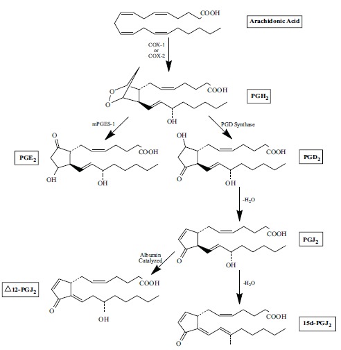
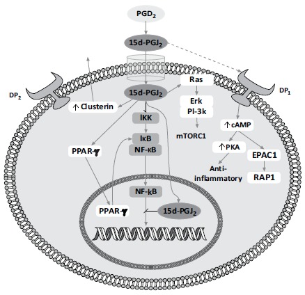
Osteoarthritis (OA) of the knee is a wide-spread, debilitating disease that is prominent in Western countries. It is associated with old age, obesity, and mechanical stress on the knee joint. By examining the recent literature on the effect of the anti-inflammatory prostaglandins 15d-PGJ2 and Δ12-PGJ2, we propose that new therapeutic agents for this disease could facilitate the transition from the COX-2-dependent pro-inflammatory synthesis of the prostaglandin PGE2 (catalyzed by mPGES-1), to the equally COX-2-dependent synthesis of the aforementioned anti-inflammatory prostaglandins. This transition could be instrumental in halting the breakdown of cartilage via matrix metalloproteinases (MMPs) and aggrecanases, as well as promoting the matrix regeneration and synthesis of cartilage by chondrocytes. Another desirable property of new OA therapeutics could involve the recruitment of mesenchymal stem cells to the damaged cartilage and bone, possibly resulting in the generation of chondrocytes, synoviocytes, and, in the case of bone, osteoblasts. Moreover, we propose that research promoting this transition from pro-inflammatory to anti-inflammatory prostaglandins could aid in the identification of new OA therapeutics.
Keywords: Cartilage; cyclo-oxygenase; inflammation; knee osteoarthritis; prostaglandins; synovial fluid.
Copyright© Bentham Science Publishers; For any queries, please email at epub@benthamscience.org.
Figures
Similar articles
-
Long-term NSAID treatment directly decreases COX-2 and mPGES-1 production in the articular cartilage of patients with osteoarthritis.Osteoarthritis Cartilage. 2008 Dec;16(12):1484-93. doi: 10.1016/j.joca.2008.04.022. Epub 2008 Jun 10. Osteoarthritis Cartilage. 2008. PMID: 18547825 Clinical Trial.
-
Mechanical stress and prostaglandin E2 synthesis in cartilage.Biorheology. 2008;45(3-4):301-20. Biorheology. 2008. PMID: 18836232
-
Contrasting effects of peroxisome-proliferator-activated receptor (PPAR)gamma agonists on membrane-associated prostaglandin E2 synthase-1 in IL-1beta-stimulated rat chondrocytes: evidence for PPARgamma-independent inhibition by 15-deoxy-Delta12,14prostaglandin J2.Arthritis Res Ther. 2005;7(6):R1325-37. doi: 10.1186/ar1830. Epub 2005 Sep 22. Arthritis Res Ther. 2005. PMID: 16277686 Free PMC article.
-
The role of cytokines in osteoarthritis pathophysiology.Biorheology. 2002;39(1-2):237-46. Biorheology. 2002. PMID: 12082286 Review.
-
Osteoarthritis of the knee and hip. Part I: aetiology and pathogenesis as a basis for pharmacotherapy.J Pharm Pharmacol. 2012 May;64(5):617-25. doi: 10.1111/j.2042-7158.2012.01458.x. Epub 2012 Feb 7. J Pharm Pharmacol. 2012. PMID: 22471357 Review.
Cited by
-
Knockdown of TRIM8 Attenuates IL-1β-induced Inflammatory Response in Osteoarthritis Chondrocytes Through the Inactivation of NF-κB Pathway.Cell Transplant. 2020 Jan-Dec;29:963689720943604. doi: 10.1177/0963689720943604. Cell Transplant. 2020. PMID: 32757662 Free PMC article.
-
Intra-articular Administration of Allogeneic Adipose Derived MSCs Reduces Pain and Lameness in Dogs With Hip Osteoarthritis: A Double Blinded, Randomized, Placebo Controlled Pilot Study.Front Vet Sci. 2020 Aug 31;7:570. doi: 10.3389/fvets.2020.00570. eCollection 2020. Front Vet Sci. 2020. PMID: 33110913 Free PMC article.
-
Metabolic phenotypes reflect patient sex and injury status: A cross-sectional analysis of human synovial fluid.Osteoarthritis Cartilage. 2024 Sep;32(9):1074-1083. doi: 10.1016/j.joca.2023.09.004. Epub 2023 Sep 15. Osteoarthritis Cartilage. 2024. PMID: 37716406
-
Biomonitoring the skeletal muscle metabolic dysfunction in knee osteoarthritis in older adults: Is Jumpstart Nutrition® Supplementation effective?Caspian J Intern Med. 2023 Fall;14(4):590-606. doi: 10.22088/cjim.14.43.590. Caspian J Intern Med. 2023. PMID: 38024172 Free PMC article. Review.
-
On the Mechanisms of Action of the Low Molecular Weight Fraction of Commercial Human Serum Albumin in Osteoarthritis.Curr Rheumatol Rev. 2019;15(3):189-200. doi: 10.2174/1573397114666181119121519. Curr Rheumatol Rev. 2019. PMID: 30451114 Free PMC article. Review.
References
-
- Jay G.D., Tantravahi U., Britt D.E., Barrach H.J., Cha C.J. Homology of lubricin and superficial zone protein (SZP): products of megakaryocyte stimulating factor (MSF) gene expression by human synovial fibroblasts and articular chondrocytes localized to chromosome 1q25. J. Orthop. Res. 2001;19(4):677–687. - PubMed
-
- Pearle A.D., Warren R.F., Rodeo S.A. Basic science of articular cartilage and osteoarthritis. Clin. Sports Med. 2005;24(1):1–12. - PubMed
-
- Kiani C., Chen L., Wu Y.J., Yee A.J., Yang B.B. Structure and function of aggrecan. Cell Res. 2002;12(1):19–32. - PubMed
-
- Prevalence of disabilities and associated health conditions among adults--United States, 1999. MMWR Morb. Mortal. Wkly. Rep. 2001;50(7):120–125. - PubMed
-
- Dillon C.F., Rasch E.K., Gu Q., Hirsch R. Prevalence of knee osteoarthritis in the United States: arthritis data from the Third National Health and Nutrition Examination Survey 1991-94. J. Rheumatol. 2006;33(11):2271–2279. - PubMed
LinkOut - more resources
Full Text Sources
Other Literature Sources
Research Materials