. 2015 Aug;6(4):449-63.
doi: 10.1007/s13244-015-0411-3. Epub 2015 May 23.

False positive and false negative diagnoses of prostate cancer at multi-parametric prostate MRI in active surveillance

Affiliations

False positive and false negative diagnoses of prostate cancer at multi-parametric prostate MRI in active surveillance

Jeffrey S Quon et al. Insights Imaging. 2015 Aug.

Abstract

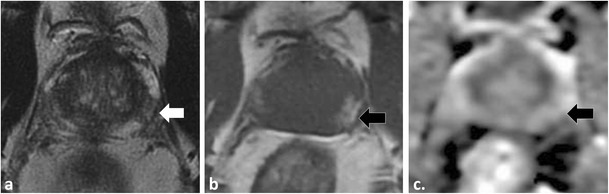
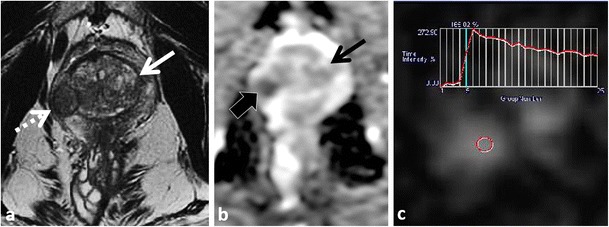
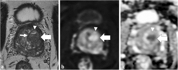
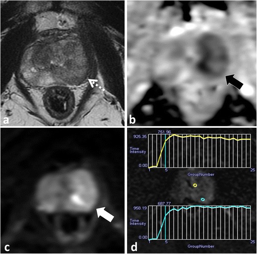
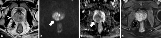
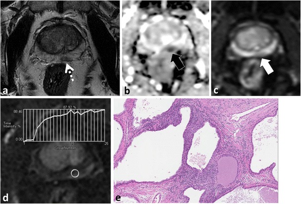
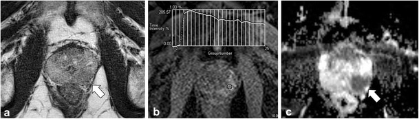
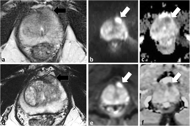
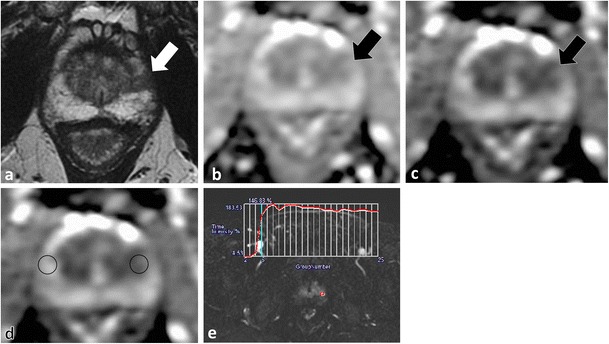
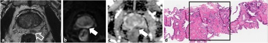
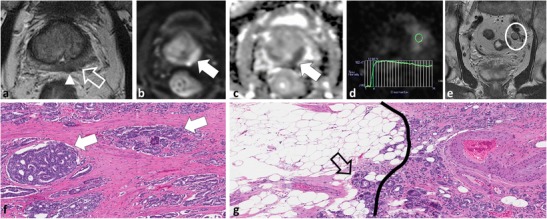
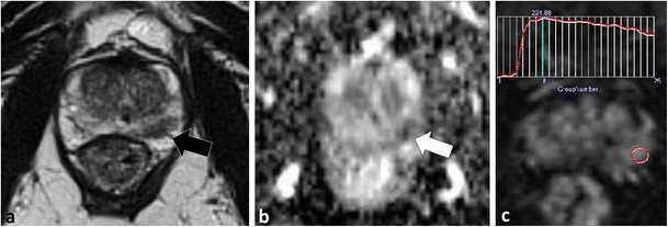
MP-MRI is a critical component in active surveillance (AS) of prostate cancer (PCa) because of a high negative predictive value for clinically significant tumours. This review illustrates pitfalls of MP-MRI and how to recognise and avoid them. The anterior fibromuscular stroma and central zone are low signal on T2W-MRI/apparent diffusion coefficient (ADC), resembling PCa. Location, progressive enhancement and low signal on b ≥1000 mm²/s echo-planar images (EPI) are differentiating features. BPH can mimic PCa. Glandular BPH shows increased T2W/ADC signal, cystic change and progressive enhancement; however, stromal BPH resembles transition zone (TZ) PCa. A rounded morphology, low T2 signal capsule and posterior/superior location favour stromal BPH. Acute/chronic prostatitis mimics PCa at MP-MRI, with differentiation mainly on clinical grounds. Visual analysis of diffusion-weighted MRI must include EPI and appropriate windowing of ADC. Quantitative ADC analysis is limited by lack of standardization; the ADC ratio and ADC histogram analysis are alternatives to mean values. DCE lacks standardisation and has limited utility in the TZ, where T2W/DWI are favoured. Targeted TRUS-guided biopsies of MR-detected lesions are challenging. Lesions detected on MP-MRI may not be perfectly targeted with TRUS and this must be considered when faced with a suspicious lesion on MP-MRI and a negative targeted TRUS biopsy histopathological result.

Keypoints: • Multi-parametric MRI plays a critical role in prostate cancer active surveillance. • Low T2W signal intensity structures appear dark on ADC, potentially simulating cancer. • Stromal BPH mimics cancer at DWI and DCE. • Long b value trace EPI should be reviewed • Targeted biopsy of MR-detected lesions using TRUS guidance may be challenging.


Figures

Fig. 1
Fig. 1
A 54-year-old male with low volume Gleason score 3 + 3 = 6 PCa at TRUS-guided biopsy in the right middle peripheral zone (PZ). MP-MRI was performed because of rising PSA to exclude clinically significant (CS) tumour. Axial (a) and coronal (b) T2-weighted (T2W) turbo spin echo (TSE) images demonstrate bilateral foci of low T2 signal intensity (SI) at the prostate base (dotted arrows) adjacent to the insertion of the vas deferens (not shown). Axial apparent diffusion coefficient (ADC) map (c) at the same level demonstrates corresponding low SI (white arrows). These areas were described as suspicious and repeat targeted TRUS-guided biopsy was suggested. A repeat biopsy with multiple cores through both areas yielded only normal prostatic tissue. A repeat MRI performed 2 years later demonstrates similar findings on the T2W (d) and ADC map (e), which represent the normal central zone. Corresponding b1000 mm²/s trace echo-planar image (EPI) does not show concordant areas of increased SI (white arrows in f) and there is benign progressive enhancement on dynamic contrast-enhanced (DCE) imaging (g)
Fig. 2
Fig. 2
A 51-year-old patient with low-volume Gleason score 3 + 3 = 6 at non-targeted TRUS-guided biopsy. MP-MRI was performed at onset of active surveillance (AS) to exclude higher grade tumour. Axial (a) and sagittal (b) T2W images demonstrate an ill-defined ovoid lesion of low T2 SI at the junction of the right PZ and transition zone (TZ) (dotted white arrow) along the course of the surgical capsule. Axial ADC map image (c) at the same level demonstrates corresponding low SI (white arrow). This area was considered suspicious for tumour. Targeted TRUS biopsy was performed and yielded only normal prostate tissue. The area did not change on follow-up MP-MRI and represents asymmetry of the normal central zone. In retrospect, corresponding b1000 mm²/s trace EPI (d) did not show increased signal (white arrow) in this area
Fig. 3
Fig. 3
A 54-year-old patient with elevated and rising PSA. Axial T2W TSE (a) demonstrates a low SI focus in the anterior midline (dotted arrow) with corresponding low SI (white arrow) on the axial ADC map (b). This area was reported as suspicious for tumour. Repeat non-targeted biopsies of the PZ revealed Gleason score 3 + 4 = 7 tumour in the right PZ (not shown) and the patient underwent radical prostatectomy (RP). The structure highlighted on MRI is the normal anterior fibromuscular stroma (AFMS); note: characteristic midline anterior location on axial T2 (a) and benign progressive enhancement on DCE (c). Also note that low SI on the ADC (b) map is due to inherently low T2 SI rather than restricted diffusion; there is a lack of increased SI on trace b1000 mm²/s EPI (d). Whole-mount histopathology image of the RP specimen (e) demonstrates the AFMS (black arrow), which is predominantly composed of smooth muscle that blends with the overlying prostate stroma
Fig. 4
Fig. 4
A 50-year-old patient with rising PSA and a typical anterior tumour involving the right side of the gland. Axial T2W TSE (a) demonstrates an amorphous, off midline, lenticular-shaped, ill-defined low T2 SI region (white arrow) with low SI (black arrow) on the ADC (b) map due to restricted diffusion (note increased SI on trace b1000 mm²/s EPI (white arrow in c) and type III contrast curve kinetics on DCE (d). A targeted TRUS biopsy was performed and confirmed Gleason score 4 + 3 = 7 tumour anteriorly. Corresponding microscopic image from TRUS biopsy (e) demonstrates Gleason pattern 4 tumour
Fig. 5
Fig. 5
A 58-year-old male patient with Gleason score 3 + 3 = 6 tumour imaged with MP-MRI because of rising PSA. On axial T2W TSE (a) there is a low SI focus in the left mid PZ (white arrow), which could represent peripheral zone cancer. Corresponding axial T1W TSE (b) demonstrates increased T1 SI (black arrow) in keeping with post-biopsy haemorrhage. Axial ADC map (c) does not reveal the lesion (black arrow), which was readily diagnosed as post-biopsy haemorrhage
Fig. 6
Fig. 6
A 47-year-old patient post-total colectomy for ulcerative colitis and elevated PSA. MP-MRI was performed to evaluate for potential tumour and to plan a biopsy. Axial T2W TSE (a) image demonstrates enlargement of the central gland (TZ) consistent with BPH. There is a well-circumscribed, round, mixed but predominantly increased T2W SI nodule with internal cystic change (white arrow) and a predominantly homogeneously low T2 SI nodule at the junction of the right middle PZ and central gland (dotted arrow). The larger nodule demonstrates T2 shine-through on the ADC map (black arrow in b) and is characteristic of glandular BPH. The other nodule demonstrates restricted diffusion (thick black arrow in b) and type III kinetics at DCE (c). A diagnosis of prostate cancer was suggested. Targeted biopsy revealed normal prostatic tissue and stromal BPH. Follow-up MRI (not shown) demonstrated no change and the PSA was stable. In retrospect, the nodule is round, well-circumscribed and demonstrates a complete low T2 SI rim (a), findings that are more in keeping with stromal BPH rather than cancer
Fig. 7
Fig. 7
A 58-year-old patient with low-volume, low Gleason score 3 + 3 = 6 tumour in the right mid-peripheral zone undergoing MRI prior to repeat biopsy because of rising PSA. Axial T2W TSE (a) image demonstrates a low T2 SI nodule in the left TZ (thick white arrow). The nodule is predominantly homogeneously of low T2W SI but demonstrates a small focus of cystic change anteriorly (arrowhead). Note that the nodule is round and demonstrates a continuous low T2W SI rim (thin white arrow). The nodule demonstrates restricted diffusion, increased SI on trace b1000 mm²/s EPI (b) and low SI on the ADC map (c) (thick white arrows). Note that cystic change demonstrates T2 shine-through (arrowhead). Imaging findings are characteristic of a mixed but predominantly stromal BPH nodule
Fig. 8
Fig. 8
Acute prostatitis in a 46-year-old patient with elevated PSA. Axial T2W TSE (a) demonstrates an amorphous area of low T2 SI (dotted arrow) in the left middle PZ with corresponding low SI on ADC map (black arrow in b) due to restricted diffusion, note increased SI on trace b1000 mm²/s EPI (white arrow in c). There is an indeterminate type II contrast curve (plateau kinetics) on DCE in (d). A differential diagnosis of prostatitis or cancer was provided, and biopsy or follow-up was suggested. The patient had typical findings of acute bacterial prostatitis clinically and was treated with antibiotics with normalisation of PSA post therapy
Fig. 9
Fig. 9
A 64-year-old patient with rising PSA and history of recurrent urothelial cell carcinoma of the urinary bladder treated with intra-vesical BCG therapy. Axial T2W image (a) demonstrates a large mass infiltrating throughout the right prostate (black arrow) and breaching the prostate capsule laterally (arrowhead) consistent with extra-prostatic spread. Axial b1000 mm²/s (b) and ADC map (c) demonstrate marked restricted diffusion (white arrows) and axial image from DCE shows marked early mass-like hyper-enhancement (type III curve was depicted and is not shown). TRUS biopsy was performed and histology was compatible with diffuse granulomatous prostatitis
Fig. 10
Fig. 10
A 64-year-old patient with Gleason score 3 + 3 = 6 in <5 % of one core in the left middle PZ underwent MRI prior to enrolment into active surveillance. Axial T2W TSE image (a) demonstrates a low T2 SI focus in the left PZ (dotted arrow) with low SI on ADC (black arrow in b). Based on these findings a diagnosis of potential higher grade tumour was suggested and a repeat biopsy was performed. Saturation biopsies through the left mid PZ revealed only normal prostatic tissue and areas of chronic prostatitis. In retrospect, trace b1000 mm²/s EPI demonstrates low SI (white arrow in c), which indicates that there is no restricted diffusion in this area. Similarly, there is a benign progressive type I enhancement curve on DCE (d), which further argues against a higher grade tumour. Corresponding histopathology slide (e) demonstrates areas of chronic inflammation
Fig. 11
Fig. 11
A 59-year-old patient with Gleason 3 + 4 = 7 cancer in the left middle PZ on TRUS biopsy underwent MP-MRI for staging. Axial T2W TSE image shows a subtle lesion in the left middle PZ (white arrow in b) with type III kinetics on DCE (b) and profound restricted diffusion on ADC map (white arrow in c). Typically with conventional acinar adenocarcinoma T2W signal intensity mirrors findings on ADC map, the discordant findings with only minimally decreased T2W signal and profound restricted diffusion can be seen in ductal variant adenocarcinoma. Final histopathology after RP was Gleason score 4 + 4 = 8 tumour with dominant ductal component
Fig. 12
Fig. 12
A 63-year-old patient with elevated PSA and previously negative non-targeted TRUS-guided biopsy with persistent clinical suspicion of prostate cancer underwent MP-MRI in two separate sessions within 3 months demonstrating the loss of contrast with BLADE/PROPELLER imaging. Axial T2W BLADE a, b1000 mm²/s EPI b and ADC map c demonstrate a suspicious focus of restricted diffusion in the left middle anterior horn of the PZ (white arrows), which is not visible on axial T2W BLADE (black arrow in a). Repeat examination performed using T2W FSE (d) reveals a T2 hypointense nodule in the same location (black arrow in d) with persistent restricted diffusion (white arrows in e and f)
Fig. 13
Fig. 13
A 55-year-old patient with low-volume Gleason score 3 + 3 = 6 tumour at TRUS biopsy in the left apical PZ underwent MRI prior to consideration for potential AS. Axial T2W TSE image (a) demonstrates a low T2 SI focus in the left apical PZ (white arrow). Axial ADC map displayed with automatic windowing/levelling shows minimal decreased SI (black arrow in b). These findings could be considered typical for a 3 + 3 = 6 cancer. With modified display of the ADC map using previously validated settings (width = 1.650 and level = 1.675 × 10–6 mm²/s) the nodule (black arrow in c) is noted to be of lower SI than initially displayed in (b). Using quantitative data, the ADC value obtained within the tumour (d) was 1.462 × 10−3 mm²/s, which would also be considered to be of low (Gleason score 6) grade using previously reported thresholds. Comparing ADC values across systems is challenging due to a lack of standardization, and an ADC ratio has been previously proposed as a better metric to compare ADC. An ADC value from the contralateral normal PZ obtained at the same level was 2.000 × 10−3 mm²/s, which yields an ADC ratio of 0.73, which would be compatible with a Gleason score ≥7 tumour based on previously published thresholds. Corresponding DCE image (e) from the same level demonstrated a focal enhancing nodule with a type III contrast curve. Based on the imaging findings, a repeat TRUS-guided biopsy was performed, which demonstrated Gleason pattern 4 in the left apical PZ
Fig. 14
Fig. 14
A 59-year-old patient with low-volume Gleason score 3 + 3 = 6 at non-targeted TRUS biopsy underwent MP-MRI to exclude clinically significant cancer after an increase in PSA. Axial T2W TSE image (a) demonstrates a low T2 SI focus in the left PZ (open white arrow) with restricted diffusion; note increased SI on trace b1000 mm²/s EPI and low SI on ADC (white arrows in b and c, respectively). Based on the MRI and clinical findings a diagnosis of potential higher grade tumour was suggested and a targeted repeat biopsy was performed. At time of repeat biopsy, which used cognitive registration of MP-MRI and TRUS data, no lesion could be identified at TRUS. Only one core biopsy through the left medial mid peripheral zone sextant was performed. Microscopic image from repeat TRUS-guided biopsy (d) demonstrates Gleason pattern 3 tumour. Continued in Fig. 14
Fig. 15
Fig. 15
Although only low-volume Gleason score 3 + 3 = 6 was again noted at repeat TRUS biopsy (Fig. 13), a follow-up MP-MRI was performed 3 months later because of interval doubling of PSA to re-evaluate for a focus of higher grade cancer. Axial T2W TSE image (a) demonstrates that the small low T2 SI focus in the left PZ has grown substantially (open white arrow) with bulging and nodular extension into the peri-prostatic fat (arrowhead), which was reported as representing extra-prostatic extension. The lesion again demonstrates restricted diffusion; note increased SI on trace b1000 mm²/s EPI and low SI on ADC (white arrows in b and c, respectively) and demonstrated an aggressive type III contrast curve on DCE (d). In the interval, a malignant-appearing lymph node developed along the left pelvic sidewall (e). Based on these findings a diagnosis of high-grade tumour with extra-prostatic extension and metastatic adenopathy was provided. The patient underwent RP based on the imaging findings, he declined a repeat biopsy because of previous urosepsis related to prior TRUS biopsy. Corresponding microscopic images (f and g) demonstrate Gleason pattern 4 tumour (white arrows in f) and extra-prostatic extension of tumour (open arrow), which is beyond the prostate capsule (black line) and into the peri-prostatic fat (g)
Fig. 16
Fig. 16
A 63-year-old patient underwent MP-MRI prior to routine repeat biopsies as part of his active surveillance protocol. Axial T2W TSE (a), axial ADC map (b) and semi-quantitative contrast curve derived from DCE (c) demonstrate a focal low T2W SI lesion (black arrow) with restricted diffusion (white arrow) and type III kinetics in the left middle peripheral zone. At repeat targeted biopsy using cognitive registration, no corresponding lesion could be identified. With a priori knowledge of the location of the lesion at MP-MRI, the TRUS operator performed three core needle biopsies through the left middle lateral and two core needle biopsies through the left middle medial PZ sextants. Results after targeted biopsies were Gleason 4 + 3 = 7 tumour with two out of three core biopsies in the left middle lateral PZ sextant

Similar articles

Cited by

References

    1. National Comprehensive Cancer Network NCCN (2012) Clinical practice guidelines in oncology: prostate cancer. Fort Washington, PA. Available via http://www.nccn.com/files/cancer-guidelines/prostate/index.html#/1. Accessed Sept. 13 2013
    1. Prostate Cancer Canada (2014) Prostate cancer Canada network. Toronto, ON Canada. Available via http://prostatecancer.ca. Accessed Sept. 2014
    1. European Association of Urology (EAU) (2012) Guidelines on prostate cancer. Arnhem, The Netherlands. Available via http://www.uroweb.org/gls/pdf/08%20Prostate%20Cancer_LR%20March%2013th%2...
    1. Thomsen FB, Brasso K, Klotz LH, Roder MA, Berg KD, Iversen P. Active surveillance for clinically localized prostate cancer–a systematic review. J Surg Oncol. 2014;109:830–835. doi: 10.1002/jso.23584. - DOI - PubMed
    1. Heidenreich A, Bellmunt J, Bolla M, et al. EAU guidelines on prostate cancer. Part 1: screening, diagnosis, and treatment of clinically localised disease. Eur Urol. 2011;59:61–71. doi: 10.1016/j.eururo.2010.10.039. - DOI - PubMed