. 2015 Aug;14(8):2111-25.
doi: 10.1074/mcp.M115.049817. Epub 2015 May 22.

Bisecting Galactose as a Feature of N-Glycans of Wild-type and Mutant Caenorhabditis elegans

Affiliations

Bisecting Galactose as a Feature of N-Glycans of Wild-type and Mutant Caenorhabditis elegans

Shi Yan et al. Mol Cell Proteomics. 2015 Aug.

Abstract

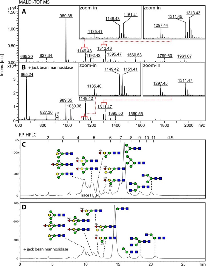
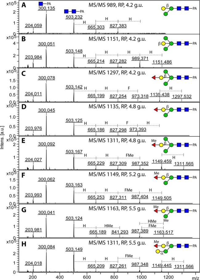
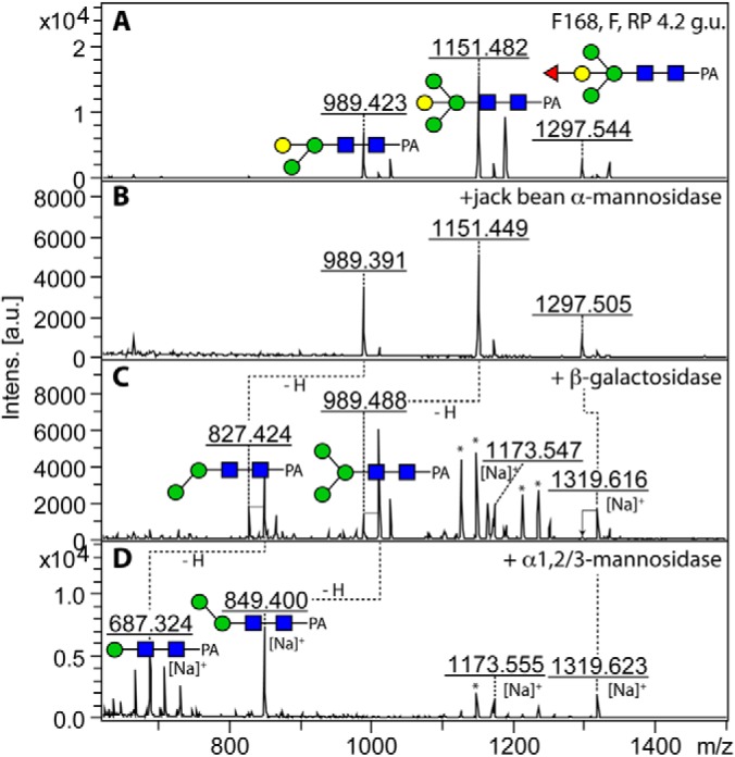
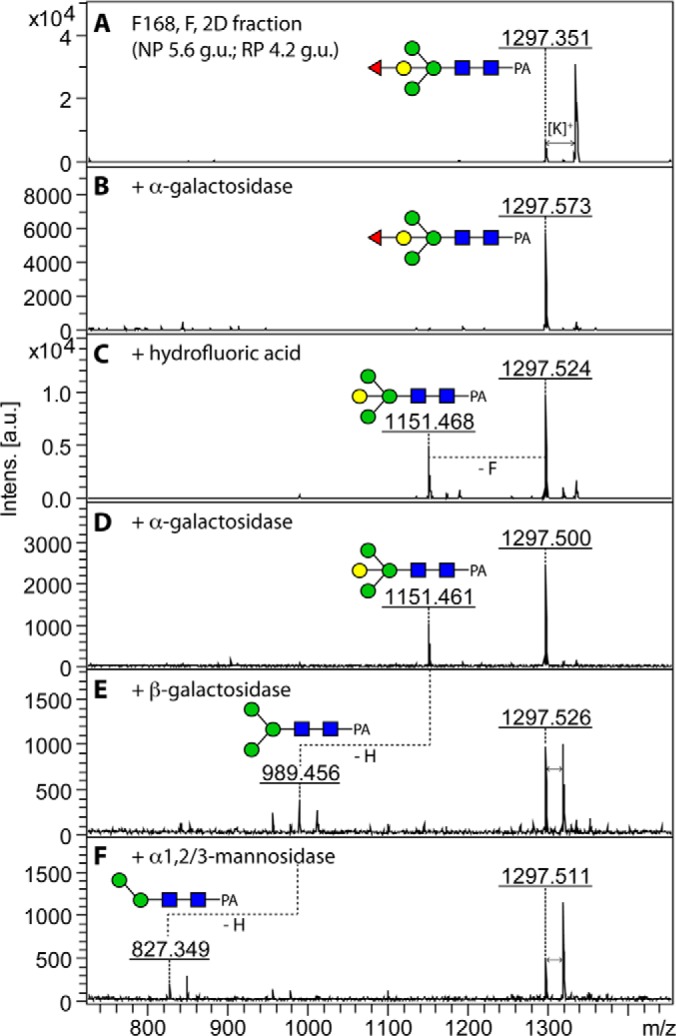
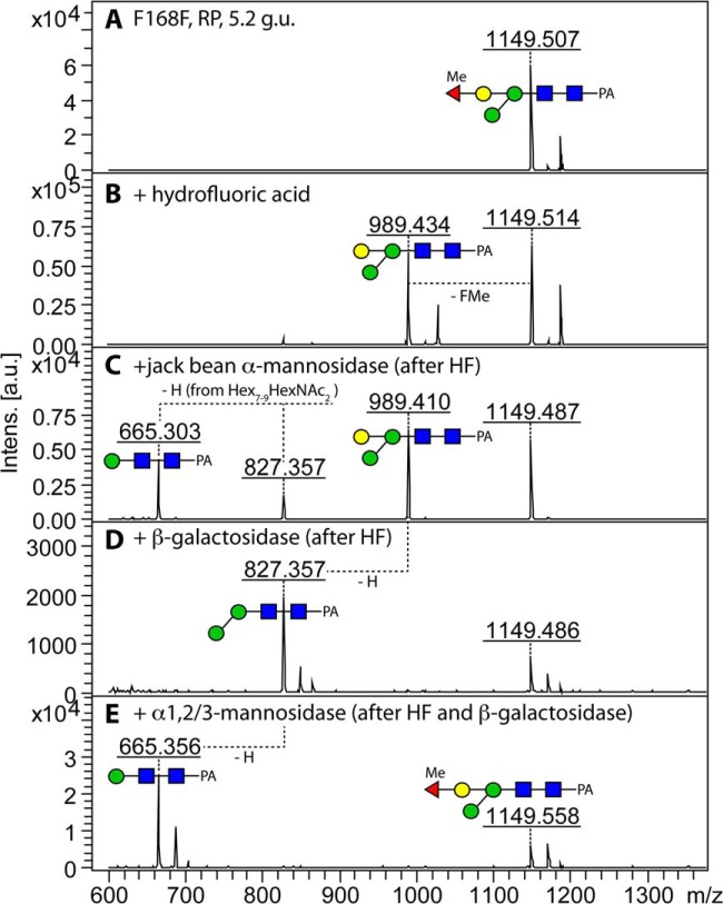
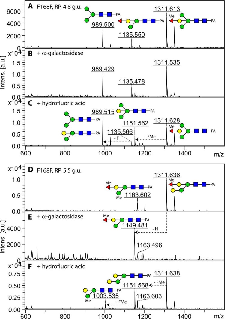
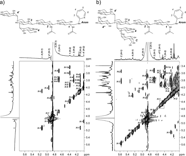
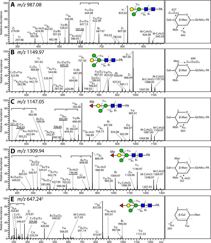
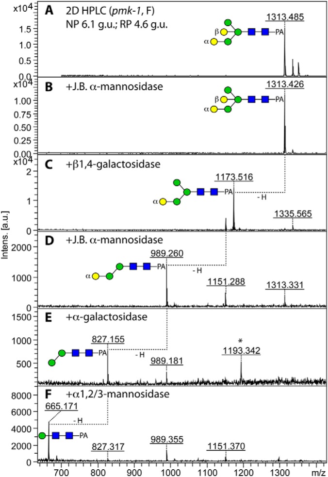
The N-glycosylation of the model nematode Caenorhabditis elegans has proven to be highly variable and rather complex; it is an example to contradict the existing impression that "simple" organisms possess also a rather simple glycomic capacity. In previous studies in a number of laboratories, N-glycans with up to four fucose residues have been detected. However, although the linkage of three fucose residues to the N,N'-diacetylchitobiosyl core has been proven by structural and enzymatic analyses, the nature of the fourth fucose has remained uncertain. By constructing a triple mutant with deletions in the three genes responsible for core fucosylation (fut-1, fut-6 and fut-8), we have produced a nematode strain lacking products of these enzymes, but still retaining maximally one fucose residue on its N-glycans. Using mass spectrometry and HPLC in conjunction with chemical and enzymatic treatments as well as NMR, we examined a set of α-mannosidase-resistant N-glycans. Within this glycomic subpool, we can reveal that the core β-mannose can be trisubstituted and so carries not only the ubiquitous α1,3- and α1,6-mannose residues, but also a "bisecting" β-galactose, which is substoichiometrically modified with fucose or methylfucose. In addition, the α1,3-mannose can also be α-galactosylated. Our data, showing the presence of novel N-glycan modifications, will enable more targeted studies to understand the biological functions and interactions of nematode glycans.


Figures

Fig. 1.
Fig. 1.
Total N-glycome of the fut-1;fut-6;fut-8 triple knockout and its sensitivity to jack bean α-mannosidase. PA-derivatised N-glycans before and after mannosidase digestion were profiled by MALDI-TOF MS (A and B) and by reversed phase HPLC (C and D); the region of panel B above m/z 900 is magnified fivefold because of the dominance of the m/z 665 mannosidase-digestion product. The major glycan structures identified in this mutant were detected as [M+H]+ and are annotated on the HPLC chromatograms according to the nomenclature of the Consortium for Functional Glycomics. The intensity is in arbitrary units (a.u.).
Fig. 2.
Fig. 2.
MALDI-TOF MS/MS spectra of pyridylamino-labeled N-glycans carrying bisecting β-galactose. N-glycans from the fut-1;fut-6;fut-8 triple mutant separated by RP-HPLC (4.2–5.5 g.u.) were subject to MS/MS. Nonfucosylated forms (A and B), fucosylated forms (C and D) and methyl-fucosylated forms (E–H) displayed sequential “loss” of hexose and (methyl-) fucose residues or methylhexose (the latter only in G). For all (methyl-) fucosylated structures, the absence of key ions such as HexNAc1Fuc1-PA (m/z 446) and Hex1HexNAc1Fuc1-PA (m/z 608) indicated that there is no core fucosylation in this triple knockout. Other key ions such as Hex2HexNAc2Me-PA (m/z 841) and Hex2HexNAc2Fuc1Me-PA (m/z 987) are indicative of the methyl group.
Fig. 3.
Fig. 3.
Sequential digestions of the RP-HPLC fraction eluting at 4.2 glucose units. Three co-eluting structures (Hex3–4HexNAc2Fuc0–1-PA, A) from the fut-1;fut-6;fut-8 (F168) triple mutant were resistant to jack bean α-mannosidase digestion (B) but sensitive to recombinant β-galactosidase (C) except for the Hex4HexNAc2Fuc1-PA structure (m/z 1297.5). The β-galactosidase products with m/z 827.4 and 989.5 were further digested by α1,2/3-mannosidase (D) and yielded respective sodiated products with m/z 687.3 and 849.4. Digestion of the m/z 1297 species is shown in Fig. 4. Asterisks indicate nonglycan contaminants.
Fig. 4.
Fig. 4.
Sequential digestions of a two-dimensional HPLC fraction containing a Hex4HexNAc2Fuc1-PA variant. A, 2D-HPLC purified form of the α-mannosidase-resistant m/z 1297 glycan shown in Fig. 3 (obtained by NP-HPLC followed by RP-HPLC) was incubated with green coffee bean α-galactosidase, but no hexose residue was removed (B); hydrofluoric acid treatment converted ca. 40% of the structure into a defucosylated form with m/z 1151.5 (C). Afterward, the product was treated with either α-galactosidase (D) or β-galactosidase; only β-galactosidase resulted in a loss of a hexose residue (E). Finally, α1,2/3-mannosidase further trimmed the β-galactosidase product and yield Hex2HexNAc2-PA with m/z 827.3 (F).
Fig. 5.
Fig. 5.
Structural analysis of a novel methylated Hex3HexNAc2Fuc1Me-PA N-glycan. This α-mannosidase resistant structure was separated by RP-HPLC, eluting at 5.2 glucose unit (A). HF treatment was first applied to the fraction and resulted in a partial conversion to Hex3HexNAc2-PA (m/z 989.4) (B). This product was incubated with either jack bean α-mannosidase or β-galactosidase, but only the latter resulted in loss of a hexose residue (C and D). Further digestion of the β-galactosidase product using α1,2/3-mannosidase resulted in removal of the “lower arm” mannose and formation of a final product with m/z 665.3.
Fig. 6.
Fig. 6.
Structural characterization of two isomeric N-glycans by α-galactosidase digestion and HF treatment. Two different RP-HPLC fractions containing Hex4HexNAc2Fuc1Me1-PA with m/z 1311.6 (4.8 g.u. and 5.5 g.u.; A and D) were treated with α-galactosidase (B and E); complete loss of one hexose was observed only for the late eluting fraction, resulting in a product of m/z 1149.5. Hydrofluoric acid treatment resulted in partial removal of either fucose or methylfucose (FMe) residues from Hex3–4HexNAc2Fuc1Me0–2-PA (C and F); thus, the fucose is not α1,3-linked (which would be fully removed by this treatment) and not α1,6-linked (which would be resistant), but the degree of release is compatible with the proposed Fucα1,2 linkage.
Fig. 7.
Fig. 7.
1H NMR and TOCSY of a standard trimannosyl N-glycan and the pool of bisected N-glycans. Separated signals in the 1H NMR spectra of the 7.2 g.u. fraction containing Man3GlcNAc2-PA (A) and of the 4.0–5.5 g.u. fractions (B, lacking the glycan of 4.8 g.u.) from the triple mutant are indicated according to the numbering shown on the structures. In the TOCSY spectra, separated cross peaks of the involved nuclei in according spin systems are marked. The chemical shifts of both compounds are listed in Table II.
Fig. 8.
Fig. 8.
Detection of fucose-substituted and nonsubstituted bisecting Gal by LC-MS/MS. LC-MS/MS spectra of N-glycans from the 4.0–5.5 g.u. fut-1;fut-6;fut-8 pool: A, a paucihexosidic N-glycan containing a bisecting galactose (Hex3HexNAc2-PA, [M-H] of m/z 987), B, a Man3GlcNAc2 N-glycan with a bisecting Gal (Hex4HexNAc2-PA, [M-H] of m/z 1150), C, an N-glycan in which a methylated Fuc is linked to the bisecting Gal (Hex3HexNAc2Fuc1Me1-PA, [M-H] of m/z 1147), (D) an bisected Man3GlcNAc2 N-glycan with a methylated fucose substitution of the bisecting Gal (Hex4HexNAc2Fuc1Me1-PA, [M-H] of m/z 1310) and E, a bisected Man3GlcNAc2 N-glycan in which Fuc is linked to the bisecting Gal (Hex4HexNAc2Fuc1-PA, [M-2H]2− of m/z 647). Schematic representations explaining the Domon and Costello nomenclature of indicative glycosidic or cross-ring cleavages are shown in the panels on the right.
Fig. 9.
Fig. 9.
Sequential enzymatic digestions of an Hex5HexNAc2 isoform. A structure (m/z 1313.5 as [M+H]+) produced by the pmk-1 strain was isolated on HPLC via a two-dimensional approach (NP-HPLC followed by RP-HPLC); this glycan elutes unusually early (4.6 g.u.) on the reversed phase column (A). Jack bean α-mannosidase digestion resulted in no loss of mannose (B), whereas β-galactosidase removed one hexose residue (C). Mannosidase digestion of the β-galactosidase product resulted in additional loss of one hexose (D) yielding a glycan (m/z 989.3), which was further trimmed down to Hex2HexNAc2-PA with green coffee bean α-galactosidase (E). Finally, the Hex2HexNAc2-PA was digested by α1,2/3-mannosidase (F) to yield the core trisaccharide structure (m/z 665.2).

Similar articles

Cited by

References

    1. Sommer R. J., Streit A. (2011) Comparative genetics and genomics of nematodes: genome structure, development, and lifestyle. Annu. Rev. Genet. 45, 1–20 - PubMed
    1. Blaxter M. (2011) Nematodes: the worm and its relatives. PLoS Biol. 9, e1001050. - PMC - PubMed
    1. Paschinger K., Gutternigg M., Rendić D., Wilson I. B. H. (2008) The N-glycosylation pattern of Caenorhabditis elegans. Carbohydr. Res. 343, 2041–2049 - PubMed
    1. Schachter H. (2009) Paucimannose N-glycans in Caenorhabditis elegans and Drosophila melanogaster. Carbohydr. Res. 344, 1391–1396 - PubMed
    1. Yazdanbakhsh M., Kremsner P. G., van Ree R. (2002) Allergy, parasites, and the hygiene hypothesis. Science 296, 490–494 - PubMed

Publication types

MeSH terms

LinkOut - more resources