

Impaired coronary and retinal vasomotor function to hyperoxia in Individuals with Type 2 diabetes
- PMID: 26002545
- PMCID: PMC5061572
- DOI: 10.1016/j.mvr.2015.05.002
Impaired coronary and retinal vasomotor function to hyperoxia in Individuals with Type 2 diabetes
Abstract
Purpose: Adults with diabetes are at a high risk of developing coronary heart disease. The purpose of this study was to assess coronary artery vascular function non-invasively in individuals with and without Type 2 diabetes and to compare these coronary responses to another microvascular bed (i.e. retina). We hypothesized that individuals with diabetes would have impaired coronary reactivity and that these impairments would be associated with impairments in retinal reactivity.
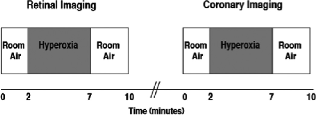
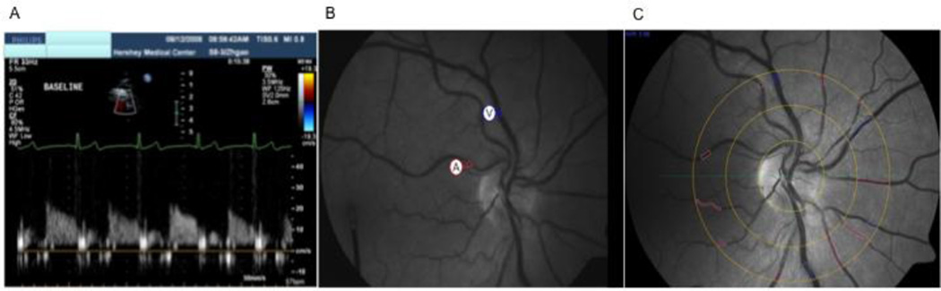
Methods: Coronary blood velocity (Transthoracic Doppler Echocardiography) and retinal diameters (Dynamic Vessel Analyzer) were measured continuously during five minutes of breathing 100% oxygen (i.e. hyperoxia) in 15 persons with Type 2 diabetes and 15 age-matched control subjects. Using fundus photographs, retinal vascular calibers were also measured (central retinal arteriole and venule equivalents).
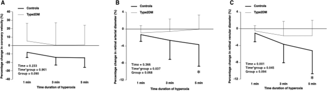
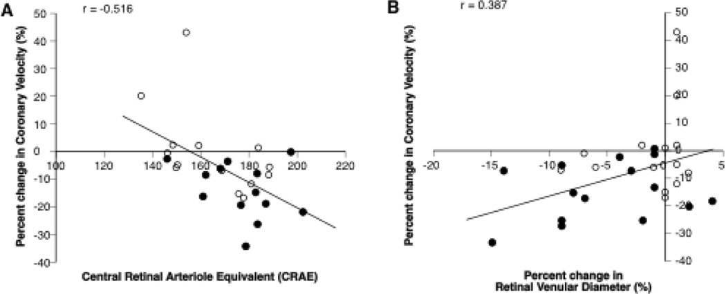
Results: Individuals with diabetes compared to controls had impaired coronary (-2.34±16.64% vs. -14.27±10.58%, P=0.03) and retinal (arteriole: -0.04±3.34% vs. -3.65±5.07%, P=0.03; venule: -1.65±3.68% vs. -5.23±5.47%, P=0.05) vasoconstrictor responses to hyperoxia, and smaller central arteriole-venule equivalent ratios (0.83±0.07 vs. 0.90±0.07, P=0.014). Coronary reactivity was associated with central retinal arteriole equivalents (r=-0.516, P=0.005) and retinal venular reactivity (r=0.387, P=0.034).
Conclusion: Diabetes impairs coronary and retinal microvascular function to hyperoxia. Impaired vasoconstrictor responses may be part of a systemic diabetic vasculopathy, which may contribute to adverse cardiovascular events in individuals with diabetes.
Keywords: Coronary reactivity; Diabetes; Hyperoxia; Retinal reactivity; Vasoconstriction.
Copyright © 2015. Published by Elsevier Inc.
Conflict of interest statement
None
Figures
Similar articles
-
Retinal arterioles have impaired reactivity to hyperoxia in type 1 diabetes.Acta Ophthalmol. 2010 Jun;88(4):453-7. doi: 10.1111/j.1755-3768.2009.01557.x. Epub 2009 Jul 21. Acta Ophthalmol. 2010. PMID: 19681793
-
Comparison of retinal vasodilator and constrictor responses in type 2 diabetes.Acta Ophthalmol. 2012 Sep;90(6):e434-41. doi: 10.1111/j.1755-3768.2012.02445.x. Epub 2012 Jun 8. Acta Ophthalmol. 2012. PMID: 22682034
-
Systemic hyperoxia and retinal vasomotor responses.Invest Ophthalmol Vis Sci. 2005 May;46(5):1714-20. doi: 10.1167/iovs.04-1216. Invest Ophthalmol Vis Sci. 2005. PMID: 15851573
-
Relative magnitude of vascular reactivity in the major arterioles of the retina.Microvasc Res. 2012 Mar;83(2):200-4. doi: 10.1016/j.mvr.2011.11.002. Epub 2011 Nov 9. Microvasc Res. 2012. PMID: 22100560 Clinical Trial.
-
The relationship between retinal microvascular abnormalities and coronary heart disease: a review.Am J Med. 2010 Apr;123(4):374.e1-7. doi: 10.1016/j.amjmed.2009.05.030. Am J Med. 2010. PMID: 20362758 Free PMC article. Review.
Cited by
-
Retinal Oxygen Metabolism in Patients With Type 2 Diabetes and Different Stages of Diabetic Retinopathy.Diabetes. 2022 Dec 1;71(12):2677-2684. doi: 10.2337/db22-0219. Diabetes. 2022. PMID: 36107468 Free PMC article.
-
A proposal for early and personalized treatment of diabetic retinopathy based on clinical pathophysiology and molecular phenotyping.Vision Res. 2017 Oct;139:153-160. doi: 10.1016/j.visres.2017.03.006. Epub 2017 Aug 2. Vision Res. 2017. PMID: 28438679 Free PMC article. Review.
-
The innate immune system in diabetic retinopathy.Prog Retin Eye Res. 2021 Sep;84:100940. doi: 10.1016/j.preteyeres.2021.100940. Epub 2021 Jan 8. Prog Retin Eye Res. 2021. PMID: 33429059 Free PMC article. Review.
-
Fractalkine-induced microglial vasoregulation occurs within the retina and is altered early in diabetic retinopathy.Proc Natl Acad Sci U S A. 2021 Dec 21;118(51):e2112561118. doi: 10.1073/pnas.2112561118. Proc Natl Acad Sci U S A. 2021. PMID: 34903661 Free PMC article.
-
Retinal vessel diameters and reactivity in diabetes mellitus and/or cardiovascular disease.Cardiovasc Diabetol. 2017 Apr 26;16(1):56. doi: 10.1186/s12933-017-0534-6. Cardiovasc Diabetol. 2017. PMID: 28446234 Free PMC article.
References
-
- Akasaka T, et al. Retinopathy identifies marked restriction of coronary flow reserve in patients with diabetes mellitus. J Am Coll Cardiol. 1997;30:935–941. - PubMed
-
- Anderson TJ, et al. Close relation of endothelial function in the human coronary and peripheral circulations. J Am Coll Cardiol. 1995;26:1235–1241. - PubMed
-
- Atar AI, et al. Coronary flow reserve in patients with diabetes mellitus and prediabetes. Echocardiography. 2012;29:634–640. - PubMed
-
- Blum M, et al. Vasoconstriction of retinal arterioles with oxygen breathing in diabetic retinopathy. Ophthalmologe. 2003;100:306–309. - PubMed
Publication types
MeSH terms
Grants and funding
LinkOut - more resources
Full Text Sources
Other Literature Sources
Medical
