. 2015 Jul 1;142(13):2364-74.
doi: 10.1242/dev.121913. Epub 2015 May 22.

The matricellular protein CCN1 controls retinal angiogenesis by targeting VEGF, Src homology 2 domain phosphatase-1 and Notch signaling

Affiliations

The matricellular protein CCN1 controls retinal angiogenesis by targeting VEGF, Src homology 2 domain phosphatase-1 and Notch signaling

Hemabindu Chintala et al. Development. .

Abstract

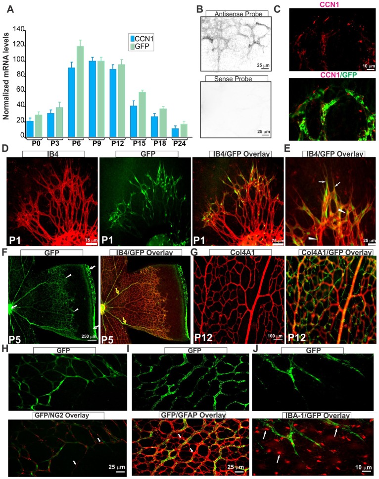
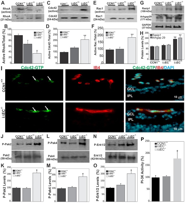
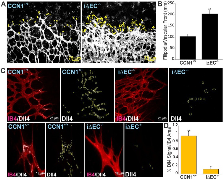
Physiological angiogenesis depends on the highly coordinated actions of multiple angiogenic regulators. CCN1 is a secreted cysteine-rich and integrin-binding matricellular protein required for proper cardiovascular development. However, our understanding of the cellular origins and activities of this molecule is incomplete. Here, we show that CCN1 is predominantly expressed in angiogenic endothelial cells (ECs) at the leading front of actively growing vessels in the mouse retina. Endothelial deletion of CCN1 in mice using a Cre-Lox system is associated with EC hyperplasia, loss of pericyte coverage and formation of dense retinal vascular networks lacking the normal hierarchical arrangement of arterioles, capillaries and venules. CCN1 is a product of an immediate-early gene that is transcriptionally induced in ECs in response to stimulation by vascular endothelial growth factor (VEGF). We found that CCN1 activity is integrated with VEGF receptor 2 (VEGF-R2) activation and downstream signaling pathways required for tubular network formation. CCN1-integrin binding increased the expression of and association between Src homology 2 domain-containing protein tyrosine phosphatase-1 (SHP-1) and VEGF-R2, which leads to rapid dephosphorylation of VEGF-R2 tyrosine, thus preventing EC hyperproliferation. Predictably, CCN1 further brings receptors/signaling molecules into proximity that are otherwise spatially separated. Furthermore, CCN1 induces integrin-dependent Notch activation in cultured ECs, and its targeted gene inactivation in vivo alters Notch-dependent vascular specification and remodeling, suggesting that functional levels of Notch signaling requires CCN1 activity. These data highlight novel functions of CCN1 as a naturally optimized molecule, fine-controlling key processes in physiological angiogenesis and safeguarding against aberrant angiogenic responses.

Keywords: CCN1; Knockout mouse; Matricellular; Retinal angiogenesis; VEGF.


Figures

Fig. 1.
Fig. 1.
CCN1 is expressed in endothelial cells in angiogenic vasculature. (A) Analysis of CCN1 and GFP transcript levels of CCN1:GFP reporter transgenic mice during postnatal development of the retinal vasculature. CCN1 and GFP mRNA levels as determined by qPCR were normalized to those of the acidic ribosomal phosphoprotein (ARBP) (n=5). (B) In situ hybridization of CCN1 sense and antisense probes in P4 flat-mounted mouse retinas, showing positive signal with the antisense probe in the retinal vasculature. (C) Immunohistochemical localization of the CCN1 protein in P4 flat-mounted retina of CCN1:GFP reporter mice. Images show CCN1 and merged CCN1/GFP signals. (D,E) Flat-mounted retinas stained with isolectin B4 (IB4; red). Images depict IB4, GFP and/or merged IB4/GFP staining. Arrows and arrowheads indicate CCN1:GFP reporter signals in endothelial tip and stalk cells, respectively. (F,G), Immunostaining of flat-mounted retinas of CCN1:GFP reporter mice at P5 and P12. Note the increasingly enriched and restricted GFP signal to the advancing vascular front (white arrowheads) and large veins (yellow arrows). GFP signal can also be seen in the remnants of hyaloid vessels found around the optic nerve and at the edge of retinal leaflets (white arrows). In G, note the absence of CCN1:GFP signal in the superficial capillaries in the plexus visualized with anti-α1 type IV collagen (Col4A1) antibodies and increased CCN1:GFP signal in deeper plexuses. (H-J) Flat-mounted CCN1:GFP mouse retinas stained with anti-NG2 (H), anti-GFAP (I) and anti-IBA-1 (J) antibodies, depicting pericyte, astrocyte and microglia localization, respectively (white arrows).
Fig. 2.
Fig. 2.
Retinal vascular abnormalities following EC-specific inactivation of the CCN1 gene. (A,B) Representative immunofluorescence images of whole-mounts of retinas of CCN1+/+, Cdh5-Cre and iΔEC−/− mice upon induction of EC-specific CCN1 deletion from P1 to P3 and analysis at P4. Staining was performed with either IB4 (a, b, c) or anti-Col4A1 antibody (d), which also allows visualization of the retinal vasculature. (C-E) Analysis and quantification of vascular parameters of representative retinas from CCN1+/+ and iΔEC−/− mice at P4 using the AngioTool software. Fields of view at the sprouting vascular front of the retinal vascular networks from control and mutant mice included regions of capillary-sized vessels directly adjacent to radial arterioles. Graphical representation of the analysis of the vascular area, branching index, lacunarity and vascular progression are shown in C, D, E and F, respectively. **P<0.01, ***P<0.001 versus CCN1+/+ (n=4-5).
Fig. 3.
Fig. 3.
Loss of CCN1 induces EC hyperproliferation. (A) Immunofluorescence images showing BrdU incorporation (green) together with IB4 staining (red) in the retina at P6 of CCN1+/+, iΔEC+/− and iΔEC−/− mice. (B) Quantitative analysis of EC proliferation at P6 as measured by counting BrdU+ nuclei. Equivalent areas of retinas from CCN1+/+, iΔEC+/− and iΔEC−/− mice were compared. Data are means±s.e.m. *P<0.05 versus CCN1+/+; **P<0.001 versus CCN1+/+ (n=5). (C) Whole-mount α1 type IV collagen (Col4A1, red) and isolectin B4 (IB4, green) staining of P5 retinas from CCN1+/+ control and iΔEC−/− mice. (D) Quantitation of empty collagen IV sleeves in iΔEC−/− and littermate control retinal vasculature. *P<0.05 versus CCN1+/+ (n=5).
Fig. 4.
Fig. 4.
Loss of CCN1 potentiates VEGF receptor activation. (A,B) Quantification by qPCR of endogenous levels of VEGF-A, VEGF-B and VEGF-C (A) and of VEGF-R1, VEGF-R2 and VEGF-R3 (B) mRNAs in CCN1+/+, iΔEC+/− and iΔEC−/− mouse retinas. Transcript levels in non-mutant CCN1+/+ were set to 100% to facilitate comparisons and analyses among the three groups of mice. *P<0.05 versus CCN1+/+ (n=5). (C,D) VEGF-R2 activation upon endothelial-specific CCN1 deletion in mice. Lysates from retinas were analyzed by immunoprecipitation with an anti-VEGF-R2 antibody followed by immunoblotting with either anti-pY1175 or anti-pY1214 antibody. VEGF-R2 protein band in the total protein input was visualized with anti-VEGF-R2 antibody. Densitometric quantification of the phosphorylated and non-phosphorylated forms of VEGF-R2 using ImageJ software is shown in D. **P<0.05 versus CCN1+/+ (n=3).
Fig. 5.
Fig. 5.
CCN1-induced SHP-1 expression/activity modulates VEGF-induced EC proliferation. (A,B) ECs were exposed to VEGF for increasing periods of time (A, left panel) in the presence or absence of a scrambled siRNA (siR-Scrbl) or siR-CCN1 (A, right panel). Protein lysates were analyzed by western immunoblotting. The proliferation rate was determined using the CyQUANT direct cell proliferation assay. *P<0.001 versus control; **P<0.005 versus VEGF+siR-Scrbl (n=3). (C) Cells were transduced with adenoviral vector expressing either GFP (Ad-GFP), VEGF (Ad-VEGF) or CCN1 (Ad-CCN1), and the proliferation rate was determined following cell transduction with either Ad-GFP, Ad-VEGF or Ad-CCN1 in serum-free medium for 16 h. *P<0.001 versus Ad-GFP; **P<0.05 versus Ad-VEGF (n=4). (D) VEGF-R2 phosphorylation at the Y1175 and Y1214 sites in cells treated as described in A,B. Cell lysates were analyzed by immunoprecipitation with an anti-VEGFR-2 antibody to determine total VEGF-R2 protein input. (E) SHP-1 and SHP-2 mRNA levels were determined by qPCR in cells transduced with either Ad-GFP or Ad-CCN1 and incubated for 16 h in serum-free medium. *P<0.001 versus Ad-GFP (n=3). (F,G) SHP-1 protein expression (F) and activity (G) in cells transfected with siR-Scrbl or siR-CCN1 and left untreated (Ctrl) or treated with VEGF. (H) Cells were transduced with Ad-CCN1 and/or Ad-VEGF and protein extracts were analyzed by western immunoblotting (upper panel). Immunoprecipitation of protein extracts with anti-SHP-1, and further detection with antibodies against VEGF-R2 or CCN1 is shown (lower panel). (I) Effect of function-blocking antibodies against different integrin subunits on the formation of the complex including VEGF-R2, SHP-1 and CCN1. Cells were incubated in serum-free medium with integrin-blocking antibodies for up to 4 h. Cell lysates were analyzed as described in H. (J-L) SHP-1 protein levels as determined by western blotting (J), densitometric scanning (K) and activity (L) in retinal lysates of CCN1+/+, iΔEC+/− and iΔEC−/− mice. **P<0.01 versus CCN1+/+ (n=3).
Fig. 6.
Fig. 6.
Loss of CCN1 potentiates VEGF-R2 downstream signaling through Rho GTAse and MAPK activation during retinal vessel formation. (A-F) Rho A, Cdc42 and Rac activation status as determined by GTPase assay in retinal extracts from wild-type, iΔEC+/− and iΔEC−/− mice. Protein band signals were normalized to total input of each GTPase (B,D,F). **P<0.01 versus CCN1+/+ (n=3). (G,H) Expression of signaling kinases (Rasip1 and Arhgap29) upstream of Cdc42/Rac1 GTPases. The same blots were stripped and washed before subsequent incubation with antibody against the indicated proteins. Experiments were performed on at least three different retinal lysate preparations with similar results. **P<0.05 versus CCN1+/+. (I) Effects of CCN1 deletion on the activation of Cdc42. Transverse sections of P6 retinas were labeled with either Cdc42-GTP-specific antibody or IB4. Note that the active Cdc42-GTP was largely localized within the vasculature of both control CCN1+/+ and iΔEC−/− mice (arrows). GCL, ganglion cell layer; IPL, inner plexiform layer. (J-O) Phosphorylation status of signaling kinases (Pak2, Pak4 and Erk1/2) downstream of Cdc42/Rac1 GTPases. Phosphorylated protein levels were normalized to those of the corresponding non-phosphorylated protein signal. *P<0.01 versus CCN1+/+. (P) PI3-K activity in retinal protein lysates as determined by PI3-K activity ELISA assay. *P<0.001 versus CCN1+/+ (n=4).
Fig. 7.
Fig. 7.
Endothelial-specific deletion of CCN1 impairs Dll4/Notch Signaling. (A) Typical vascular fronts of flat-mounted IB4-stained retinas from control CCN1+/+ and iΔEC−/− mouse pups. IB4 staining was overexposed to visualize the filopodia (scored with yellow dots) at the vascular front. (B) Quantification of filopodia-rich endothelial tip cells in the retina of iΔEC−/− mice and their control counterparts. Values represent means±s.d. Tip cells were counted in four equivalent areas of retinas of six control and six iΔEC−/− mouse retinas. *P<0.05 versus CCN1+/+. (C) Typical pattern of Dll4 protein localization in tip and stalk cells in the retina of P4 control and mutant iΔEC−/− mice. Whole-retinal-mount immunostaining for IB4 (in red) and Dll4 (white) in retinas of P4 control and mutant iΔEC−/− mice. Yellow circles highlight the Dll4 staining. (D) Quantification of the percentage of Dll4+ signal normalized to IB4+ area shown in C.

References

    1. Bautch V. L. (2012). VEGF-directed blood vessel patterning: from cells to organism. Cold Spring Harb. Perspect. Med. 2, a006452 10.1101/cshperspect.a006452 - DOI - PMC - PubMed
    1. Benedito R., Rocha S. F., Woeste M., Zamykal M., Radtke F., Casanovas O., Duarte A., Pytowski B. and Adams R. H. (2012). Notch-dependent VEGFR3 upregulation allows angiogenesis without VEGF–VEGFR2 signalling. Nature 484, 110-114. 10.1038/nature10908 - DOI - PubMed
    1. Bhattacharya R., Kwon J., Wang E., Mukherjee P. and Mukhopadhyay D. (2008). Src homology 2 (SH2) domain containing protein tyrosine phosphatase-1 (SHP-1) dephosphorylates VEGF Receptor-2 and attenuates endothelial DNA synthesis, but not migration. J Mol. Signal. 3, 8 10.1186/1750-2187-3-8 - DOI - PMC - PubMed
    1. Brigstock D. R. (2003). The CCN family: a new stimulus package. J. Endocrinol. 178, 169-175. 10.1677/joe.0.1780169 - DOI - PubMed
    1. Chang K.-H., Chan-Ling T., McFarland E. L., Afzal A., Pan H., Baxter L. C., Shaw L. C., Caballero S., Sengupta N., Calzi S. L. et al. (2007). IGF binding protein-3 regulates hematopoietic stem cell and endothelial precursor cell function during vascular development. Proc. Natl. Acad. Sci. USA 104, 10595-10600. 10.1073/pnas.0702072104 - DOI - PMC - PubMed

Publication types

MeSH terms

Substances

LinkOut - more resources