. 2015 Aug:80:70-9.
doi: 10.1016/j.nbd.2015.04.016. Epub 2015 May 21.

Inflammatory mechanisms contribute to the neurological manifestations of tuberous sclerosis complex

Affiliations

Inflammatory mechanisms contribute to the neurological manifestations of tuberous sclerosis complex

Bo Zhang et al. Neurobiol Dis. 2015 Aug.

Abstract

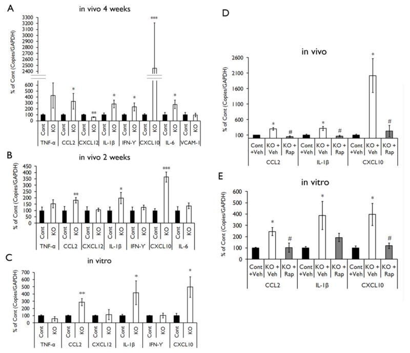
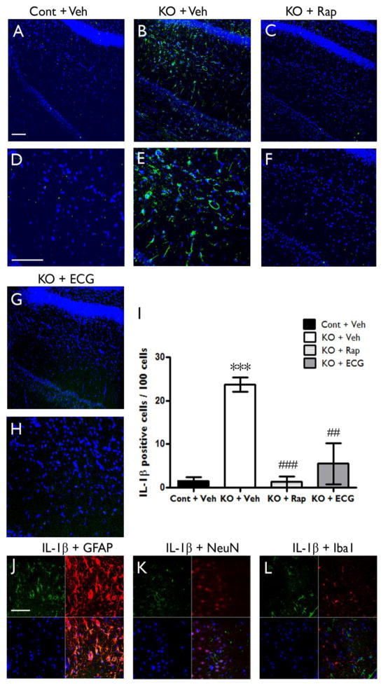
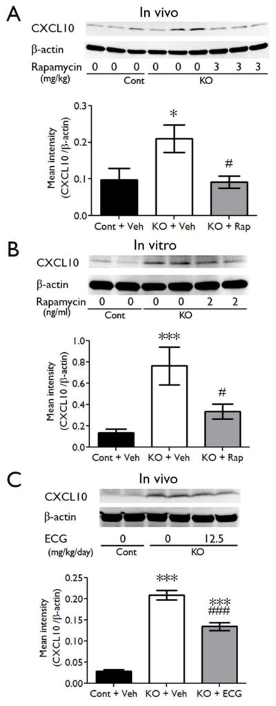
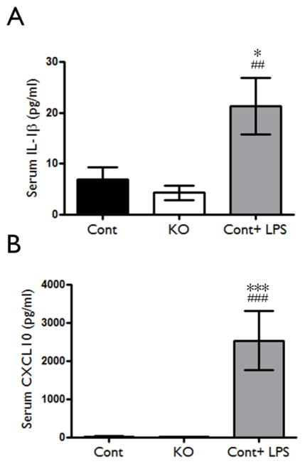
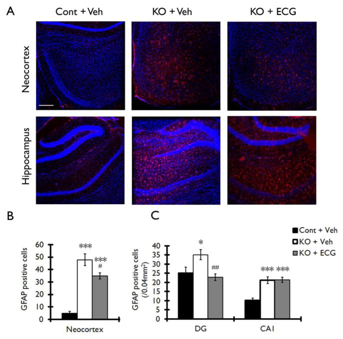
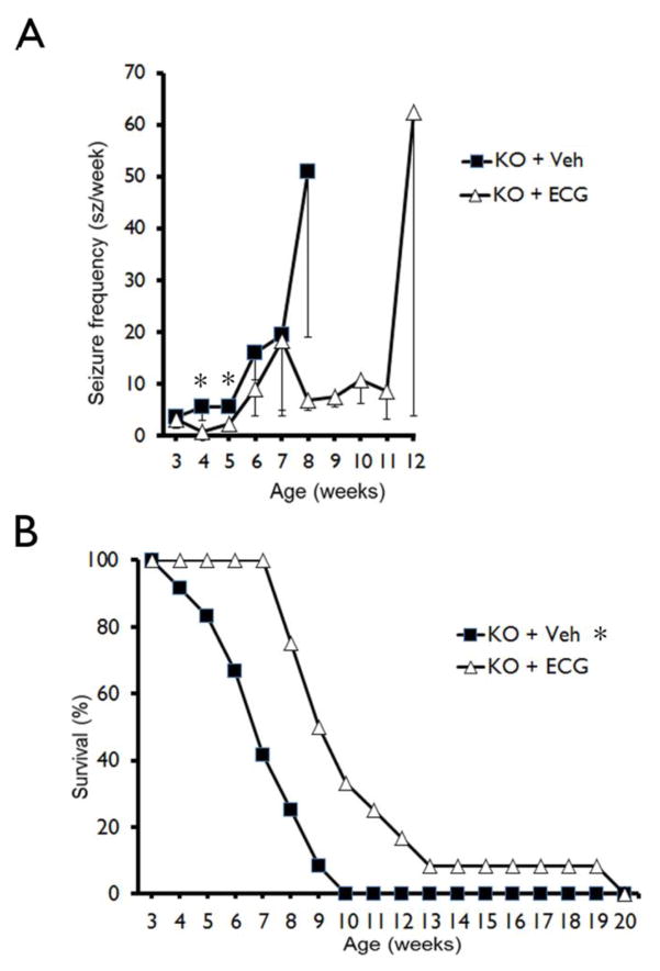
Epilepsy and other neurological deficits are common, disabling manifestations of the genetic disorder, tuberous sclerosis complex (TSC). Brain inflammation has been implicated in contributing to epileptogenesis in acquired epilepsy due to brain injury, but the potential role of inflammatory mechanisms in genetic epilepsies is relatively unexplored. In this study, we investigated activation of inflammatory mediators and tested the effects of anti-inflammatory treatment on epilepsy in the Tsc1-GFAP conditional knock-out mouse model of TSC (Tsc1(GFAP)CKO mice). Real-time quantitative RT-PCR, immunohistochemistry, and Western blotting demonstrated increased expression of specific cytokines and chemokines, particularly IL-1β and CXCL10, in the neocortex and hippocampus of Tsc1(GFAP)CKO mice, which was reversed by treatment with a mammalian target of rapamycin complex 1 (mTORC1) inhibitor. Double-labeling immunohistochemical studies indicated that the increased IL-1β was localized primarily to astrocytes. Importantly, the increase in inflammatory markers was also observed in astrocyte culture in vitro and at 2 weeks of age in Tsc1(GFAP)CKO mice before the onset of epilepsy in vivo, indicating that the inflammatory changes were not secondary to seizures. Epicatechin-3-gallate, an inhibitor of IL-1β and CXCL10, at least partially reversed the elevated cytokine and chemokine levels, reduced seizure frequency, and prolonged survival of Tsc1(GFAP)CKO mice. These findings suggest that mTOR-mediated inflammatory mechanisms may be involved in epileptogenesis in the genetic epilepsy, TSC.

Keywords: Chemokine; Cytokine; Epilepsy; Inflammation; Interleukin; Mice; Seizure; Tuberous sclerosis.


Figures

Figure 1
Figure 1. Proinflammatory cytokines and chemokines are up-regulated in Tsc1GFAPCKO mice and inhibited by rapamycin
mRNA expression of cytokines and chemokines was evaluated by real-time quantitative RT-PCR in the brains or cultured astrocytes from Tsc1GFAPCKO and control mice. (A) The mRNA levels of CCL2, IL-1β, IFN-γ, CXCL10 and IL-6 were increased in four-week old Tsc1GFAPCKO mice compared with control mice, but CXCL12 was decreased (n = 8–10 mice/group). (B) The mRNA levels of CCL2, IL-1β and CXCL10 were increased in two-week old Tsc1GFAPCKO mice compared with control mice (n = 8–11 mice/group). (C) The mRNA levels of CCL2, IL-1β and CXCL10 were increased in cultured astrocytes from Tsc1GFAPCKO mice compared with astrocytes from control mice (n = 5–9 mice/group). * p < 0.05, ** p < 0.01, *** p < 0.001, versus control mice by Student’s t test. (D) Seven days of rapamycin treatment (3 mg/kg/d i.p.) significantly inhibited the mRNA levels of CCL2, IL-1β and CXCL10 in the brains of Tsc1GFAPCKO mice compared with vehicle-treated Tsc1GFAPCKO mice (n = 4–12 mice/group). (E) Rapamycin treatment (2 ng/ml for 16 hours) significantly inhibited the mRNA levels of CCL2 and CXCL10, but not IL-1β, in cultured astrocytes from Tsc1GFAPCKO mice (n = 5–12 mice/group). * p<0.05 versus control mice, # p < 0.05 versus vehicle-treated Tsc1GFAPCKO mice by one-way ANOVA. Cont = control mice, KO = Tsc1GFAPCKO, Veh = vehicle, Rap = Rapamycin.
Figure 2
Figure 2. IL-1β protein is upregulated in Tsc1GFAPCKO mice and inhibited by anti-inflammatory treatments
Protein expression of IL-1β was assessed by immunohistochemistry in Tsc1GFAPCKO and control mice, as well as in rapamycin or ECG treated Tsc1GFAPCKO mice. (A–H) Confocal images of immunohistochemical staining of IL-1β (green) and TO-PRO-3 Iodide (blue). TO-PRO-3 Iodide (blue) was used as an optimal fluorescence dye for nuclear counterstaining. IL-1β protein expression was detected in brain sections of Tsc1GFAPCKO mice (B,E KO + Veh), but not in control mice (A,D Cont + Veh). Rapamycin (C,F KO + Rap) and ECG (G,H KO + ECG) treatment inhibited the IL-1β expression. Scale bars = 100 μm. (I) Quantitative analysis confirmed an increase in IL-1β-positive cells (per 100 TO-PRO-3 positive cells) in vehicle-treated Tsc1GFAPCKO group (KO + Veh) compared with vehicle-treated control group (Cont + Veh). Rapamycin (3 mg/kg/d i.p. for one week) or ECG treatment (12.5 mg/kg/d i.p. for one week) significantly decreased IL-1β-positive cells. ***p<0.05 versus vehicle-treated control mice (n=4–5 mice/group); ## p<0.01, ### p<0.001 versus vehicle-treated Tsc1GFAPCKO mice by two-way ANOVA. (J, K, L) Double label immunofluorescence confocal microscopy for expression of IL-1β protein (green) with GFAP (F, red for astrocytes), NeuN (G, red for neurons) and Iba1 (H, red for microglia) within brain sections of four-week-old Tsc1GFAPCKO mice. All sections were also labeled with TO-PRO-3 Iodide (blue) as an optimal fluorescence dye for nuclear counterstaining. IL-1β was found co-localized with GFAP, but not with NeuN and Iba1. Scale bar = 50 μm. Cont = control, KO = Tsc1GFAPCKO, TO PRO3 = TO-PRO-3 Iodide, ECG = Epicatechin-3-gallate, Rap = Rapamycin.
Figure 3
Figure 3. CXCL10 protein is upregulated in Tsc1GFAPCKO mice and reversed by anti-inflammatory treatment in vivo and in vitro
Protein expression of CXCL10 was assessed by western blotting in the brains and cultured astrocytes of Tsc1GFAPCKO mice, and the effects of rapamycin and ECG were tested. (A) Vehicle-treated Tsc1GFAPCKO mice have significantly increased CXCL10 levels, compared with control mice. Rapamycin treatment (3 mg/kg/d i.p. for seven days) significantly inhibited the upregulated-CXCL10 in Tsc1GFAPCKO mice (7–11 mice/group). (B) Vehicle-treated cultured astrocytes from Tsc1GFAPCKO mice showed increased CXCL10 expression compared with astrocytes from control mice. Rapamycin treatment (2 ng/ml added to the culture medium for 16 hours) blocked the up-regulation of CXCL10 in Tsc1GFAPCKO astrocyte (n=8–12 mice/group). (C) Vehicle-treated Tsc1GFAPCKO mice have significantly increased CXCL10 protein expression compared with control mice. ECG treatment (12.5 mg/kg/d i.p. for seven days) inhibited the increased CXCL10 in Tsc1GFAPCKO mice, but this was still significantly higher than control mice (n=8 mice/group). * p<0.05, *** p<0.001, versus vehicle-treated control mice or astrocytes by one-way ANOVA; # p<0.05, ### p<0.001, versus vehicle-treated Tsc1GFAPCKO mice or astrocytes by one-way ANOVA (n = 9–12 mice/group). Cont = control, KO = Tsc1GFAPCKO, Veh = vehicle, Rap = Rapamycin, ECG = Epicatechin-3-gallate.
Figure 4
Figure 4. Lack of evidence for systemic inflammation in Tsc1GFAPCKO mice
Serum inflammatory markers were assessed in Tsc1GFAPCKO mice to evaluate the potential involvement of the peripheral immune system in contributing to brain inflammation. There was no significant difference in serum IL-1β (A) and CXCL10 (B) protein levels as assayed by ELISA in 4 week-old Tsc1GFAPCKO mice compared with control mice, while LPS induced a significant increase in these markers in control mice. * p<0.05, *** p<0.001, versus vehicle-treated control mice by one-way ANOVA; ## p<0.01, ### p<0.001, versus vehicle-treated Tsc1GFAPCKO mice by one-way ANOVA (n = 5–9 mice/group). Cont = control, KO = Tsc1GFAPCKO.
Figure 5
Figure 5. ECG treatment decreases the number of GFAP positive cells in neocortex and hippocampus of Tsc1GFAPCKO mice
The effect of ECG on the number of GFAP positive cells was assessed in Tsc1GFAPCKO mice by immunohistochemistry. (A) Vehicle-treated Tsc1GFAPCKO mice (KO + Veh) displayed a diffuse increase in GFAP-positive cells (red) in neocortex (upper middle panel) and hippocampus (lower middle panel) compared with the vehicle-treated control mice (Cont + Veh). ECG treatment partially prevented this increase in GFAP-positive cells in Tsc1GFAPCKO mice (KO + ECG). TO-PRO-3 Iodide (blue) was used as an optimal fluorescence dye for nuclear counterstaining. (B, C) Quantitative analysis confirmed an increase in GFAP-positive cells in vehicle-treated Tsc1GFAPCKO group (KO + Veh) compared with vehicle-treated control group (Cont + Veh) in neocortex, dentate gyrus (DG) and CA1 of hippocampus. ECG treatment (12.5 mg/kg/d i.p. for four weeks) decreased GFAP-positive cells in neocortex and DG, but not in CA1 (KO + ECG; n=8 mice/group). *p<0.05, *** p<0.001 versus vehicle-treated control mice by two-way ANOVA; #p<0.05, ## p<0.01 versus vehicle-treated Tsc1GFAPCKO mice by two-way ANOVA. Scale bar = 200 μm. Cont = control mice, KO = Tsc1GFAPCKO mice, Veh = vehicle, ECG = Epicatechin-3-gallate, DG = dentate gyrus, CA1 = CA1 pyramidal cell layer of hippocampus.
Figure 6
Figure 6. ECG treatment does not prevent neuronal disorganization in Tsc1GFAPCKO mice
The effect of ECG on neuronal organization was assessed in Tsc1GFAPCKO mice by cresyl violet staining. Compared with control mice (A), vehicle-treated Tsc1GFAPCKO mice (B) exhibited widely dispersed pyramidal cell layers (arrows) in all regions of hippocampus (CA1–CA4). ECG treated Tsc1GFAPCKO mice (C) had a similar pattern as vehicle-treated Tsc1GFAPCKO group (B), with no apparent effect on this neuronal disorganization. Scale bar = 500 μm. Cont = control mice, KO = Tsc1GFAPCKO mice, Veh = vehicle, ECG = Epicatechin-3-gallate.
Figure 7
Figure 7. ECG treatment slightly decreases the development of seizures and improves survival in Tsc1GFAPCKO mice
(A) Seizures start to develop in vehicle-treated Tsc1GFAPCKO mice (A, KO + Veh) around 3 weeks and become progressively more frequent with age. ECG treatment (12.5 mg/kg/d i.p. starting at 3 weeks of age (KO + ECG) slightly decreased seizure frequency in Tsc1GFAPCKO mice at 4 and 5 weeks of age compared with vehicle-treated Tsc1GFAPCKO mice. (*p<0.05 by one-way ANOVA, n = 12 mice/group). However, all mice were found to have at least one seizure in both vehicle-treated and ECG-treated Tsc1GFAPCKO mice. (B) Survival analysis showed that vehicle-treated Tsc1GFAPCKO mice die prematurely with 50% mortality between 6–7 weeks of age and 100% mortality by 10 weeks. ECG treatment (12.5 mg/kg/d i.p. starting at 3 weeks of age) significantly improved the survival of Tsc1GFAPCKO mice compared to vehicle treated Tsc1GFAPCKO mice, but all ECG-treated mice still died by 20 weeks of age. *p<0.05 by Chi-Square test, comparing the two groups (n = 12 mice/group). KO = Tsc1GFAPCKO, Veh = vehicle, ECG = Epicatechin-3-gallate.

Similar articles

Cited by

References

    1. Bargerstock E, Puvenna V, Iffland P, Falcone T, Hossain M, Vetter S, Man S, Dickstein L, Marchi N, Ghosh C, Carvalho-Tavares J, Janigro D. Is peripheral immunity regulated by blood-brain barrier permeability changes? PLoS One. 2014;9:e101477. - PMC - PubMed
    1. Berdichevsky Y, Dryer AM, Saponijian Y, Mahoney MM, Pimentel CA, Lucini CA, Usenovic M, Staley KJ. PI3K-Akt signaling activates mTOR-mediated epileptogenesis in organotypic hippocampal culture model of post-traumatic epilepsy. J Neurosci. 2013;33:9056–9067. - PMC - PubMed
    1. Boer K, Jansen F, Nellist M, Redeker S, van den Ouweland AM, Spliet WG, van Nieuwenhuizen O, Troost D, Crino PB, Aronica E. Inflammatory processes in cortical tubers and subependymal giant cell tumors of tuberous sclerosis complex. Epilepsy Res. 2008;78:7–21. - PubMed
    1. Boer K, Crino PB, Gorter JA, Nellist M, Jansen FE, Spliet WG, van Rijen PC, Wittink FR, Breit TM, Troost D, Wadman WJ, Aronica E. Gene expression analysis of tuberous sclerosis complex cortical tubers reveals increased expression of adhesion and inflammatory factors. Brain Pathol. 2010;20:704–719. - PMC - PubMed
    1. Chu-Shore CJ, Major P, Camposano S, Muzykewicz D, Thiele E. The natural history of epilepsy in tuberous sclerosis complex. Epilepsia. 2010;51:1236–1241. - PMC - PubMed

Publication types

MeSH terms