. 2015 Nov;35(8):1217-26.
doi: 10.1007/s10571-015-0214-6. Epub 2015 May 24.

Expression of Peroxiredoxin 1 After Traumatic Spinal Cord Injury in Rats

Affiliations

Expression of Peroxiredoxin 1 After Traumatic Spinal Cord Injury in Rats

Shen Huang et al. Cell Mol Neurobiol. 2015 Nov.

Abstract

Reactive astrogliosis and microgliosis after spinal cord injury (SCI) contribute to glial scar formation that impedes axonal regeneration. The mechanisms underlying reactive astrocyte and microglia proliferation upon injury remain partially understood. Peroxiredoxin 1 (PRDX1) is an antioxidant participating in cell proliferation, differentiation, and apoptosis. However, PRDX1 functions in SCI-induced astrocyte and microglia proliferation are unknown. In this study, we established an acute spinal cord contusion injury model in adult rats to investigate the potential role of PRDX1 during the pathological process of SCI. We found the palpable expression increase of PRDX1 after SCI by western blot and immunohistochemistry staining. Double immunofluorescence staining showed that PRDX1 expression mainly increased in astrocytes and microglia. In addition, PRDX1/proliferating cell nuclear antigen (PCNA) colocalized in astrocytes and microglia. Furthermore, PCNA expression also elevated after SCI, as well as was positively correlated with PRDX1 expression. In vitro, PRDX1 expression in primary rat spinal cord astrocytes and microglia changed in a concentration- and time-dependent manner according to LPS treatment. In addition, PRDX1 knockdown in astrocytes and microglia resulted in the decrease of PCNA expression after LPS stimulation, showing that PRDX1 promoted astrocyte and microglia proliferation after inflammation. Our results suggested that PRDX1 might play a crucial role in astrocyte and microglia proliferation after SCI.

Keywords: Glial cell proliferation; Peroxiredoxin 1 (PRDX1); Rats; Spinal cord injury (SCI).


Figures

Fig. 1
Fig. 1
The hind limb locomotor score of rats on the BBB scale at each time point after injury
Fig. 2
Fig. 2
PRDX1 expression increased after SCI. a Sample immunoblots probed PRDX1 and GAPDH. b Quantification of the relative densitometry protein levels for PRDX1 relative to GAPDH. *P<0.05
Fig. 3
Fig. 3
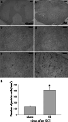
PRDX1 expression and distribution in rat spinal cord. Low-power views of spinal cord cross-sections immunostained with antibody to PRDX1 in sham (a) and 5 days injury groups (b). Higher-power views in the ventral horn of gray matter (c, d) and white matter (e, f). The number of PRDX1 positive cells in sham and 5 days injury groups (g).*P<0.05. Scale bars 200 μm (a, b) and 20 μm (cf)
Fig. 4
Fig. 4
PRDX1 was expressed in astrocytes and microglia of rat spinal cord. i, l, o, r The co-expression of PRDX1 with GFAP or CD11b. af Double stain for PRDX1 and NeuN. s The ratio of GFAP or CD11b positive cells expressing PRDX1 in sham and 5 days injury groups. *, # P < 0.05. Scale bars 20 μm (a–r)
Fig. 5
Fig. 5
PRDX1 was associated with astrocyte and microglia proliferation after SCI. PCNA expression in rat spinal cord (a, b). The colocalization of PCNA and GFAP (ot), PCNA and CD11b (in). in, rt The PCNA positive cells in rat spinal cord at 5 days after SCI. fh The colocalization of PRDX1 and PCNA (ch). The colocalization of PRDX1 GFAP and PCNA (ux). Scale bars 20 μm
Fig. 6
Fig. 6
PRDX1 promoted astrocyte proliferation after LPS stimulation. PCNA was detected in LPS-induced astrocytes with different concentrations (a, b). PCNA expression in 1.0 μg/ml LPS-treated astrocytes for 1, 3, 6, 12, or 24 h (c, d). Three different siRNAs were used to inhibit PRDX1 expression in astrocytes (e, f). The most effective PRDX1 siRNA1 was applied to detect PRDX1 and PCNA expression in astrocytes with or without LPS treatment (g, h)
Fig. 7
Fig. 7
PRDX1 promoted microglia proliferation after LPS stimulation. PCNA was detected in LPS-induced microglia with different concentrations (a, b). PCNA expression in 10 μg/ml LPS-treated microglia for 1, 3, 6, 12, or 24 h (c, d). The most effective PRDX1 siRNA1 was applied to detect PRDX1 and PCNA expression in microglia with or without LPS treatment (e, f)

References

    1. Bao Y, Qin L, Kim E, Bhosle S, Guo H, Febbraio M, Haskew-Layton RE, Ratan R, Cho S (2012) CD36 is involved in astrocyte activation and astroglial scar formation. J Cereb Blood Flow Metab 32(8):1567–1577 - PMC - PubMed
    1. Basso DM, Beattie MS, Bresnahan JC (1995) A sensitive and reliable locomotor rating scale for open field testing in rats. J Neurotrauma 12(1):1–21 - PubMed
    1. Becher B, Prat A, Antel JP (2000) Brain-immune connection: immuno-regulatory properties of CNS-resident cells. Glia 29(4):293–304 - PubMed
    1. Castriotta RJ, Murthy JN (2009) Hypoventilation after spinal cord injury. Semin Respir Crit Care Med 30(3):330–338 - PubMed
    1. Fitch MT, Silver J (2008) CNS injury, glial scars, and inflammation: inhibitory extracellular matrices and regeneration failure. Exp Neurol 209(2):294–301 - PMC - PubMed

LinkOut - more resources