Comparative Study
. 2015 Jun 2;23(6):1116-22.
doi: 10.1016/j.str.2015.03.025. Epub 2015 May 21.

NMR Structure of Francisella tularensis Virulence Determinant Reveals Structural Homology to Bet v1 Allergen Proteins

Affiliations
Comparative Study

NMR Structure of Francisella tularensis Virulence Determinant Reveals Structural Homology to Bet v1 Allergen Proteins

James Zook et al. Structure. .

Abstract

Tularemia is a potentially fatal bacterial infection caused by Francisella tularensis, and is endemic to North America and many parts of northern Europe and Asia. The outer membrane lipoprotein, Flpp3, has been identified as a virulence determinant as well as a potential subunit template for vaccine development. Here we present the first structure for the soluble domain of Flpp3 from the highly infectious Type A SCHU S4 strain, derived through high-resolution solution nuclear magnetic resonance (NMR) spectroscopy; the first structure of a lipoprotein from the genus Francisella. The Flpp3 structure demonstrates a globular protein with an electrostatically polarized surface containing an internal cavity-a putative binding site based on the structurally homologous Bet v1 protein family of allergens. NMR-based relaxation studies suggest loop regions that potentially modulate access to the internal cavity. The Flpp3 structure may add to the understanding of F. tularensis virulence and contribute to the development of effective vaccines.


Figures

Figure 1
Figure 1
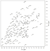
Assigned 2D 15N-HSQC spectrum of Flpp3sol. NMR spectrum displays a wide proton dispersion with narrow peak widths, indicative of a protein with significant beta sheet and alpha helix content.
Figure 2
Figure 2
Structure of Flpp3sol and model of Flpp3 inserted into the membrane. (A) Overlay of the 20 lowest energy models after water bath refinement. (B) Cartoon model of the lowest energy model of Flpp3sol. Residues F37, S41, A61, D63, and S66 may be dynamically important as per initial relaxation results and are shown in violet. (C and D) The transmembrane helix (residues Met1 to Thr25) modelled onto the N-terminus of Flpp3sol and inserted into a POPC/POPE membrane. MD production simulations were run for 1 ns with experimental restraints followed by another 1 ns without restraints. The hydrophobic region of the helix spans the entire length of the membrane with a positively charged N-terminus (not seen). Model is rotated 180° to show both sides of the protein. An electrostatic surface is plotted onto the model to show polarity between the membrane proximal region (blue/positive) and the membrane distal region (red/negative).
Figure 3
Figure 3
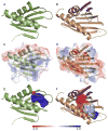
Comparison of Flpp3sol and structurally homologous Bet v1 protein birch pollen allergen (1FM4). (A and B) Secondary structure and overall fold topology of Flpp3sol (A) and 1FM4 (B). The Bet v1 proteins are larger due to two extra beta strands that distort the first alpha helix (purple). (C and D) Electrostatic surface plotted onto Flpp3sol (C) and 1FM4 (D). Flpp3sol has a polarized surface with a negatively charged (red) side and a positively charged (blue) region compared to 1FM4. (E and F) The internal cavity is shown for Flpp3sol (E) and 1FM4 (F). The two extra beta strands (purple) in 1FM4 provide additional cavity space that is limited by Flpp3sol.
Figure 4
Figure 4
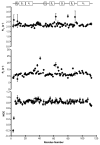
15N NMR relaxation studies. R1 data does not vary considerably between residues and demonstrates an overall constant rate of 2.2 ± 0.2 s−1. R2 data reveals a fairly constant relaxation rate throughout the structured regions of Flpp3sol. Flexible termini show slightly decreased values while several residues in the loop region reveal much greater R2 relaxation rates which may be the result of chemical exchange. In the steady-state heteronuclear NOE experiment, NOE values are relatively large and unchanging. The histidine tail did show a negative NOE values but was left out of the analysis due to inability to resolve individual histidine residues.

Similar articles

Cited by

References

    1. Baker NA, Sept D, Joseph S, Holst MJ, McCammon JA. Electrostatics of nanosystems: Application to microtubules and the ribosome. Proc Natl Acad Sci U S A. 2001;98:10037–10041. - PMC - PubMed
    1. Bhattacharya A, Tejero R, Montelione GT. Evaluating protein structures determined by structural genomics consortia. Proteins. 2007;66:778–795. - PubMed
    1. Bieri M, d’Auvergne EJ, Gooley PR. relaxGUI: a new software for fast and simple NMR relaxation data analysis and calculation of ps-ns and mu s motion of proteins. J Biomol NMR. 2011;50:147–155. - PubMed
    1. Cai CZ, Han LY, Ji ZL, Chen X, Chen YZ. SVM-Prot: web-based support vector machine software for functional classification of a protein from its primary sequence. Nucleic Acids Res. 2003;31:3692–3697. - PMC - PubMed
    1. Case DA, Cheatham TE, Darden T, Gohlke H, Luo R, Merz KM, Onufriev A, Simmerling C, Wang B, Woods RJ. The Amber biomolecular simulation programs. J Comput Chem. 2005;26:1668–1688. - PMC - PubMed

Publication types

Associated data

LinkOut - more resources