. 2015 Jun 22;33(6):690-702.
doi: 10.1016/j.devcel.2015.04.009. Epub 2015 May 21.

Slitrk5 Mediates BDNF-Dependent TrkB Receptor Trafficking and Signaling

Affiliations

Slitrk5 Mediates BDNF-Dependent TrkB Receptor Trafficking and Signaling

Minseok Song et al. Dev Cell. .

Abstract

Recent studies in humans and in genetic mouse models have identified Slit- and NTRK-like family (Slitrks) as candidate genes for neuropsychiatric disorders. All Slitrk isotypes are highly expressed in the CNS, where they mediate neurite outgrowth, synaptogenesis, and neuronal survival. However, the molecular mechanisms underlying these functions are not known. Here, we report that Slitrk5 modulates brain-derived neurotrophic factor (BDNF)-dependent biological responses through direct interaction with TrkB receptors. Under basal conditions, Slitrk5 interacts primarily with a transsynaptic binding partner, protein tyrosine phosphatase δ (PTPδ); however, upon BDNF stimulation, Slitrk5 shifts to cis-interactions with TrkB. In the absence of Slitrk5, TrkB has a reduced rate of ligand-dependent recycling and altered responsiveness to BDNF treatment. Structured illumination microscopy revealed that Slitrk5 mediates optimal targeting of TrkB receptors to Rab11-positive recycling endosomes through recruitment of a Rab11 effector protein, Rab11-FIP3. Thus, Slitrk5 acts as a TrkB co-receptor that mediates its BDNF-dependent trafficking and signaling.


Conflict of interest statement

Competing Financial Interests

The authors declare no competing financial interests.

Figures

Figure 1
Figure 1. TrkB receptors interact and co-localize with Slitrk5
(A) Interaction between TrkB receptors and Slitrk5 was assessed in HEK293T overexpressing cDNAs encoding FLAG-TrkB, Slitrk5, and empty vector. Cell lysates were immunoprecipitated with anti-FLAG antibodies and immunoblotted with anti-Slitrk5 antibodies. (B) Interaction between TrkB receptors and Slitrk5 was assessed in HEK293-TrkB cells. Lysates from Slitrk5-GFP or empty vector transfected HEK293-TrkB cells were immunoprecipitated with anti-GFP antibodies and immunoblotted with anti-TrkB antibody. (C) Endogenous association of Slitrk5 and TrkB. Mouse whole-brain lysates (2 months old) were subjected to immunoprecipitation with anti-TrkB antibody (Millipore) or control IgG. The immune protein complex was then eluted, and TrkB and Slitrk5 were detected by immunoblotting. (D) and (E) Interaction between TrkB and Slitrk5 is specific. (D) Interaction between TrkB and Slitrk family members. FLAG-tagged Slitrk family isotypes were expressed in HEK293-TrkB cells. Cell lysates were immunoprecipitated with anti-TrkB (Millipore) antibodies and immunoblotted with anti-FLAG (M2) antibodies (E) Dissociated E16 mouse cortical neurons were electroporated (Amaxa) with FLAG-TrkB or FLAG-TrkC. At DIV6, cell lysates were precipitated with anti-FLAG (M2) antibodies and immunoblotted with anti-Slitrk5 antibodies. (F) First LRR domain of Slitrk5 mediates TrkB binding. HEK293-TrkB cells were transfected with FLAG-tagged WT Slitrk5, chimeric FLAG-LRR1-domain-swapped (FLAG-LRR1(S1) Slitrk5) and LRR2-domain-swapped Slitrk5 (FLAG-LRR2(S1) Slitrk5) or empty vector. Cell lysates were precipitated with anti-FLAG (M2) antibodies and immunoblotted with anti-TrkB (Millipore) antibodies. Schematic representation of chimeric Slitrk5 mutants shown on the right. (G) LRR domain of TrkB mediates Slitrk5 binding. Dissociated E16 mouse cortical neurons were transfected with FLAG-tagged WT TrkB, LRR domain-swapped TrkB (FLAG-LRR(C) TrkB), and IgG1 domain-swapped TrkB (FLAG-IgG1(C) TrkB) or empty vector. At DIV6, cell lysates were immunoprecipitated with anti-FLAG antibodies and immunoblotted with anti-Slitrk5. Schematic representation of chimeric TrkB mutants shown on the right. (H) BDNF-dependent and TrkB kinase activity-dependent binding of TrkB and Slitrk5. After transfection of FLAG-tagged Slitrk5 into HEK293-TrkB cells, cells were pretreated with DMSO or K252a for 30 min to inhibit kinase activity of TrkB after overnight serum starvation. Cells were treated with or without BDNF, and their binding was examined by immunoprecipitation with anti-TrkB antibodies followed by immunoblotting with anti-FLAG antibodies. (I) TrkB receptors co-localize with Slitrk5 in a BDNF-dependent manner. DIV6 striatal neurons were treated with or without BDNF (25ng/ml) for 30 min after incubating with anti-TrkB antibody to specifically label surface TrkB. Endogenous Slitrk5 and MAP2 were visualized with specific antibodies after fixation and permeabilization. Super resolution images were acquired using a Nikon N-SIM structured illumination microscope. The co-localization of TrkB and Slitrk5 was presented using co-localization highlighter (ImageJ) and 3D reconstruction (IMARIS) to show more convincing co-localization signals. (J) Histogram showing mean Manders’ coefficients for co-localization of TrkB and Slitrk5 (n=20 for each condition). High Manders’ coefficients indicate better co-localization of TrkB with Slitrk5. Results are means ± SEM from 3 independent experiments. 20–30 neurons were analyzed per condition per experiment. *P<0.05 significantly different from control condition (Student’s t test).
Figure 2
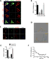
Figure 2. PTPδ and TrkB compete for binding to Slitrk5
(A) Slitrk5 binds PTPδ through LRR1. Soluble purified PTPδ-Fc chimeras were added to HEK293T cells expressing WT and indicated deletion mutants of Slitrk5 and the binding analyzed by immunofluorescence microscopy. Note that the observed binding to WT Slitrk5 is abolished by deletion of Slitrk5’s extracellular domain (ECD) or LRR1 but not LRR2. (B) Quantitative analysis of these results. Results are means ± SEM from 3 independent experiments. 20–30 cells were analyzed per condition per experiment. ***P< 0.0001 significantly different from WT condition. (One-way ANOVA and Dunnett’s multiple comparisons test). (C) BDNF displaces Slitrk5 binding from PTPδ to TrkB. Heterophilic cell adhesion assay in which HEK293-TrkB cells expressing FLAG-Slitrk5 are co-cultured with HEK293T cells expressing HA-PTPδ. Surface proteins were visualized by fluorescence microscopy in the presence or absence of BDNF treatment (as described in Supplemental Experimental Procedures). (D) Fluorescence intensity trace of FLAG-Slitrk5 (green), HA-PTPδ (red), and TrkB (blue) scanning across corresponding purple arrow as indicated in (C). (E) Quantitative colocalization analysis of FLAG-Slitrk5-HA-PTPδ and FLAG-Slitrk5-TrkB in the presence or absence of BDNF treatment in (C). Results are means ± SEM from 3 independent experiments. 10–15 cells showing heterophilic adhesion were analyzed per condition per experiment. ***P< 0.0001 significantly different from control condition. (2-way ANOVA and Sidak’s multiple comparisons test). (F) BDNF-induced dissociation of pre-bound PTPδ-Fc from HA-Slitrk5-expressing HEK293-TrkB cells. HA-Slitrk5-expressing HEK293-TrkB cells were pre-incubated with saturating condition of PTPδ-Fc (400 nM) for 1hr. After washing, cells were incubated with indicated dose of BDNF for 30 min. Remaining PTPδ-Fc binding was analyzed by immunofluorescence microscopy. Results are means ± SEM from 3 independent experiments. 20–30 cells were analyzed per condition per experiment.
Figure 3
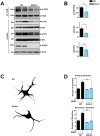
Figure 3. Altered TrkB receptor activation and signaling in the striatum of Slitrk5−/− mice
(A) Representative blots showing effect of loss of Slitrk5 on TrkB receptor activation and its downstream signaling. Striatal lysates from WT and Slitrk5−/− mice (3 months old, n=5 for each genotype) were used for immunoblot analysis for phospho-TrkB, phospho-Akt and phospho-Erk, with respective loading controls. (B) Densitometric quantification of the results shown on the right. Results are means ± SEM from 3 independent experiments. (C) and (D) Effects of BDNF on the growth of WT and Slitrk5−/− striatal neurons. Cultured striatal neurons from WT and Slitrk5−/− mice were treated with or without BDNF (40 ng/ml) at DIV2. After 5 days exposure of BDNF, the cultures were fixed and stained with anti-GAD65/67 antibody. Neuronal processes were counted with fluorescent microscopy. Representative images of BDNF-treated WT and Slitrk5−/− striatal neuron were shown in (C). Quantitation of the number of primary and secondary dendrites in WT and Slitrk5−/− was shown in (D). Results are presented as means ± SEM from 3 independent experiments determined from analysis of 40 neurons per condition per experiment (***P < 0.0001, Student’s t test).
Figure 4
Figure 4. Slitrk5 plays pivotal role in ligand dependent TrkB receptor recycling
(A) Representative blot showing enhanced BDNF-induced TrkB degradation in Slitrk5−/− striatal neurons. WT and Slitrk5−/− striatal neurons were transduced with HA-tagged WT Slitrk5, or chimeric HA-tagged-LRR1-domain-swapped (HA-LRR1(S1) Slitrk5)-expressing, or empty vector lentivirus at DIV2. Neurons were surface biotinylated and incubated at 37°C for 90 min in the absence or presence of 25 ng/ml BDNF at DIV6. Surface-labeled receptors were detected by streptavidin pull-down followed by anti-TrkB immunoblotting. (B) Densitometric quantification of the results from 3 independent experiments shown in (A) (*P < 0.05, Student’s t test). (C) and (D) TrkB recycling was impaired in Slitrk5−/− striatal neurons. TrkB recycling was examined in WT and Slitrk5−/− neurons with live-cell fluorescence ratiometric recycling assay. WT and Slitrk5−/− striatal neurons were co-transfected with FLAG-tagged TrkB lentivirus, HA-tagged WT Slitrk5 or chimeric HA-tagged-LRR1-domain-swapped (HA-LRR1(S1) Slitrk5)-expressing, or empty vector lentivirus at DIV2. Internalization of TrkB receptor was induced by BDNF treatment for 30 min after labeling surface FLAG-tagged TrkB with Alexa-488 dye-conjugated anti-FLAG (M1) antibody. Remaining surface anti-FLAG (M1) antibodies were removed with EDTA-containing Ca2+/Mg2+ free PBS, and then recycling of FLAG-tagged TrkB were monitored in the presence of Cy3-conjugated anti-mouse secondary antibody in culture medium. (C) Representative images from FLAG-tagged TrkB recycling experiment in striatal neurons. Control refers to the 100% surface TrkB receptor control, Strip refers to the 0% recycled control. The right panels of each images showed enlarged images of framed regions. (D) Receptor recycling was quantitated as described in Experimental Procedures. Results are presented as means ± SEM from 3 independent experiments determined from analysis of 30 neurons per condition per experiment (*** P <0.001, *P < 0.05, Student’s t test).
Figure 5
Figure 5. Slitrk5 facilitates TrkB receptor recruitment into Rab11-positive compartments
(A) Representative images showing co-localization of TrkB receptors and Slitrk5 in Rab11-positive compartments. WT striatal neurons were transduced with HA-tagged Slitrk5 lentivirus at DIV2. Live neurons were incubated with anti-TrkB and anti-HA antibody to specifically label cell surface proteins, and then stimulated with or without BDNF for 30 min at DIV6. Rab11 was stained after fixation and permeablization. Super resolution images were acquired using a Nikon N-SIM structured illumination microscope. (B) Representative images showing requirement of Slitrk5 for TrkB localization into Rab11-positive compartments after BDNF treatment. Co-localization of TrkB receptors and Rab11 was examined with WT and Slitrk5−/− striatal neurons in the presence or absence of BDNF. Lower panels showed enlarged images of numbered regions in upper panels. (C) Co-localization of TrkB and Rab11 was quantitated as described in Experimental Procedure. Results are presented as means ± SEM from 3 independent experiments determined from analysis of n ≥ 30 neurons per condition per experiment (*** P <0.001, Student’s t test).
Figure 6
Figure 6. Slitrk5 interacts with Rab11-FIP3 to facilitate TrkB receptor trafficking to Rab11-positive recycling endosomes
(A) and (B). Representative blots showing specific interaction between Slitrk5 and Rab11-FIP3. (A) HEK293T cells were transfected with cDNAs encoding FLAG-Slitrk5, and HA-Rab11-FIP3. Cell lysates were immunoprecipitated with anti-HA antibodies and immunoblotted with anti-FLAG antibodies. (B) HEK293T cells were transfected with cDNAs encoding FLAG-Slitrk5, and either empty vector, Rab11-FIP3-GFP, FIP1C-GFP, FIP4-GFP or FIP5-GFP. Cell lysates were immunoprecipitated with anti-GFP antibodies and blotted with anti-FLAG antibodies. (C) Representative image showing the co-localization of TrkB and Slitrk5 with Rab11-FIP3 with BDNF-dependent manner. WT striatal neurons expressing HA-Slitrk5 were stimulated with or without BDNF after incubating with anti-TrkB and anti-HA antibody for surface protein labeling. Neurons were stained with anti-Rab11-FIP3 antibody after fixation and permeablization. Super resolution images were acquired using a Nikon N-SIM structured illumination microscope. (D) Representative images showing requirement of Slitrk5 for TrkB receptor localization in Rab11-FIP3 compartments. Co-localization of TrkB and Rab11-FIP3 was examined with WT and Slitrk5−/− striatal neurons in the presence or absence of BDNF. Lower panels showed enlarged images of numbered regions in upper panels. (E) Co-localization of TrkB and Rab11-FIP3 was quantitated as described in Experimental Procedure. Results are presented as means ± SEM from 3 independent experiments determined from analysis of n ≥ 20 neurons per condition per experiment (*P<0.05, ***P<0.001, Student’s t test).
Figure 7
Figure 7. Slitrk5-mediated Rab11-FIP3 recruitment is required for TrkB recycling
(A) TrkB recycling was quantified with live-cell fluorescence ratiometric recycling assay in the control siRNA (Scrb) or Rab11-FIP3 siRNA transfected striatal neuron. Knockdown efficiency of siRNA targeting Rab11-FIP3 and representative images of live-cell fluorescence ratiometric recycling assay were shown in supplementary information (Figure S4A–C). The error bars represent the SEM of three independent experiments (n ≥ 30 cells for each condition per experiment; ** P <0.001, Student’s t test). (B) ERM domain of Rab11-FIP3 mediates Slitrk5 binding. HEK293T cells were co-transfected with cDNAs encoding FLAG-Slitrk5, and either empty vector, HA-Rab11-FIP3, HA-Rab11-FIP3ΔRBD, or HA-Rab11-FIP3ΔERM. Cell lysates were immunoprecipitated with anti-HA antibodies and immunoblotted with anti-FLAG antibodies. Schematic of Rab-FIP3 denoting established domains (EF Hand, ERM (ezrin/radixin/moesin domain), RBD (Rab11 binding domain). (C) Mapping domain in Slitrk5 that mediates association with Rab11-FIP3. Schematic representation shows a series of Slitrk5 deletion mutants that were tested for the capacity to interact with Rab11-FIP3 (Left). HEK293T cells were co-transfected with cDNAs encoding HA-Rab11-FIP3 and either FLAG-Slitrk5 or FLAG-Slitrk5ΔFIP3BD. Cell lysates were immunoprecipitated with anti-FLAG antibodies and immunoblotted with anti-HA antibodies (Right). (D) Rab11-FIP3 binding is required for Slitrk5 to rescue reduced recycling of TrkB in Slitrk5−/− striatal neurons. WT and Slitrk5−/− striatal neurons were co-transfected at DIV2 with FLAG-tagged TrkB lentivirus, and either empty vector, HA-Slitrk5 or HA-tagged Rab11-FIP3 binding-deficient Slitrk5 (HA-Slitrk5ΔFIP3BD). BDNF-induced TrkB recycling was measured with live-cell fluorescence ratiometric recycling assay at DIV6, as described in Experimental Procedure. The error bars represent the SEM of three independent experiments (n ≥ 30 cells for each condition per experiment; *** P <0.0001, Student’s t test). (E) A cell surface biotinylation assay shows that RAB11-FIP3 binding-deficient Slitrk5 is not able to rescue enhanced degradation of TrkB. WT and Slitrk5−/− striatal neurons were transduced with empty vector, HA-tagged WT Slitrk5, or HA-Slitrk5ΔFIP3BD-expressing lentivirus at DIV2. At DIV6 neurons were surface biotinylated and incubated in the presence or absence of BDNF (25ng/ml; 90 min). Cell lysates were subjected to avidin pull-down, and TrkB levels assessed by immnunoblotting with anti-TrkB antibodies. (F) Densitometric quantification of the results was shown. The error bars represent the SEM of three independent experiments (*P < 0.05, Student’s t test).

Similar articles

Cited by

References

    1. Abelson JF, Kwan KY, O’Roak BJ, Baek DY, Stillman AA, Morgan TM, Mathews CA, Pauls DL, Rasin MR, Gunel M, et al. Sequence variants in SLITRK1 are associated with Tourette’s syndrome. Science. 2005;310:317–320. - PubMed
    1. Arevalo JC, Waite J, Rajagopal R, Beyna M, Chen ZY, Lee FS, Chao MV. Cell survival through Trk neurotrophin receptors is differentially regulated by ubiquitination. Neuron. 2006;50:549–559. - PubMed
    1. Aruga J, Mikoshiba K. Identification and characterization of Slitrk, a novel neuronal transmembrane protein family controlling neurite outgrowth. Molecular and cellular neurosciences. 2003;24:117–129. - PubMed
    1. Baquet ZC, Gorski JA, Jones KR. Early striatal dendrite deficits followed by neuron loss with advanced age in the absence of anterograde cortical brain-derived neurotrophic factor. The Journal of neuroscience: the official journal of the Society for Neuroscience. 2004;24:4250–4258. - PMC - PubMed
    1. Baydyuk M, Russell T, Liao GY, Zang K, An JJ, Reichardt LF, Xu B. TrkB receptor controls striatal formation by regulating the number of newborn striatal neurons. Proceedings of the National Academy of Sciences of the United States of America. 2011;108:1669–1674. - PMC - PubMed

Publication types

MeSH terms