. 2015 Jul 17;427(14):2396-409.
doi: 10.1016/j.jmb.2015.05.011. Epub 2015 May 22.

Negative Epistasis and Evolvability in TEM-1 β-Lactamase--The Thin Line between an Enzyme's Conformational Freedom and Disorder

Affiliations

Negative Epistasis and Evolvability in TEM-1 β-Lactamase--The Thin Line between an Enzyme's Conformational Freedom and Disorder

Eynat Dellus-Gur et al. J Mol Biol. .

Abstract

Epistasis is a key factor in evolution since it determines which combinations of mutations provide adaptive solutions and which mutational pathways toward these solutions are accessible by natural selection. There is growing evidence for the pervasiveness of sign epistasis--a complete reversion of mutational effects, particularly in protein evolution--yet its molecular basis remains poorly understood. We describe the structural basis of sign epistasis between G238S and R164S, two adaptive mutations in TEM-1 β-lactamase--an enzyme that endows antibiotics resistance. Separated by 10 Å, these mutations initiate two separate trajectories toward increased hydrolysis rates and resistance toward second and third-generation cephalosporins antibiotics. Both mutations allow the enzyme's active site to adopt alternative conformations and accommodate the new antibiotics. By solving the corresponding set of crystal structures, we found that R164S causes local disorder whereas G238S induces discrete conformations. When combined, the mutations in 238 and 164 induce local disorder whereby nonproductive conformations that perturb the enzyme's catalytic preorganization dominate. Specifically, Asn170 that coordinates the deacylating water molecule is misaligned, in both the free form and the inhibitor-bound double mutant. This local disorder is not restored by stabilizing global suppressor mutations and thus leads to an evolutionary cul-de-sac. Conformational dynamism therefore underlines the reshaping potential of protein's structures and functions but also limits protein evolvability because of the fragility of the interactions networks that maintain protein structures.

Keywords: conformational diversity; interactions network; protein disorder; protein evolution; protein folds.


Figures

Fig. 1
Fig. 1. Superposition of active-site loops in all structures
(A) Superposition of the main Ω-loop conformation in all wild-type structures (gray) and in the different v13-R164S mutant structures (blue). Room-temperature structures are in green; note that in the double-mutant (R164S/G238S) room temperature structure, the Ω-loop could not be modeled and is therefore missing. Shown in sticks are the side-chains of N170 that aligns the de-acylating water (W1; gray for wild-type, red for mutants) and the catalytic S70 for reference. (B) Superposition of the main 238-loops conformation in all v13 structures containing the G238S mutation (blue), in wild-type (gray). The mutants structures determined at room temperature are in green. Shown is A237 whose backbone amide positions the oxyanion-hole water (W2). The loop conformations shown relate to the main clearly observed in the electron density of individual structures. Structures included are: A 4OPY, 4OPQ, 4OP5, 4OQI, 4OQH, 4OQ0, 4OQG, 1ZG4; B 4OPQ, 4OP8, 4OP4, 4OQI, 4OPZ, 4OQ0, 4OQG, 1ZG4 (ref. 38) (supplementary Table 1).
Fig. 2
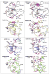
Fig. 2. The Ω-loop and 238-loop interactions network
A snapshot of the Ω-loop, the 238-loop, and the key catalytic residues in the free structures (left column) and in structures with the inhibitor EC25 bound (right column). Shown are wild-type TEM-1 (gray), the single v13 mutants R164S (yellow) and G238S (blue), and the double v13 mutant R164S/G238S (green). Catalytic residues are in smoked pink and EC25 is in light pink ball-sticks. Minor conformations of the loops are shown in light blue. Dashed lines represent putative interactions involving R164 and G238, and interactions lost in the R164S/G238S double mutant are marked in blue in the wild-type. The non-native interaction in the double mutant between E171 and S238 is in red. A putative stacking interaction between R164 and R178 (ref. 71) is also marked.
Fig. 3
Fig. 3. Electron density maps of the 238 and Ω-loops
The wild-type structure is shown for comparison in partially transparent magenta. The 2mFo-DFc electron density maps for the two v13 single mutants, and the v13 double mutant, are contoured at 0.5 (cyan) and 1 (blue) sigma, and mFo-DFc electron density maps are contoured at +3.0 (green) and −3.0 (red) sigma. (The m and D terms indicate that the maps were calculated while correcting for down-weight poorly phased reflections, and including a scaling factor to account for overall scattering differences due to missing components such as partially ordered waters).
Fig. 4
Fig. 4. The mutations impair TEM-1’s catalytic pre-organization
Superposition of the key active-sites residues in wild-type (gray), the v13 single G238S (blue) and R164S (yellow) mutants, and the v13 double mutant R164S/G238S (green). (A) Ligand-free structures. (B) The EC25 inhibitor complexes (EC25 is shown in ball-sticks). In the free structures, W1 represents the de-acylating water and W2 in the oxyanion-hole water. In the EC25-bound structures, the borate’s oxygen atoms, marked as O1 and O2, sit in the locations of W1 and W2, respectively. Note the dual conformation of N170 in the EC25 complex of R164S/G238S.

Similar articles

Cited by

References

    1. Breen MS, Kemena C, Vlasov PK, Notredame C, Kondrashov FA. Epistasis as the primary factor in molecular evolution. Nature. 490:535–8. - PubMed
    1. de Visser JA, Krug J. Empirical fitness landscapes and the predictability of evolution. Nat Rev Genet. 2014;15:480–90. - PubMed
    1. Weinreich DM, Delaney NF, Depristo MA, Hartl DL. Darwinian evolution can follow only very few mutational paths to fitter proteins. Science. 2006;312:111–4. - PubMed
    1. Bridgham JT, Ortlund EA, Thornton JW. An epistatic ratchet constrains the direction of glucocorticoid receptor evolution. Nature. 2009;461:515–9. - PMC - PubMed
    1. Borman AM, Paulous S, Clavel F. Resistance of human immunodeficiency virus type 1 to protease inhibitors: selection of resistance mutations in the presence and absence of the drug. J Gen Virol. 1996;77(Pt 3):419–26. - PubMed

Publication types