. 2015 Oct;29(7):1369-76.
doi: 10.1016/j.tiv.2015.05.008. Epub 2015 May 21.

Role of GDF15 (growth and differentiation factor 15) in pulmonary oxygen toxicity

Affiliations

Role of GDF15 (growth and differentiation factor 15) in pulmonary oxygen toxicity

Kirti Kumar Tiwari et al. Toxicol In Vitro. 2015 Oct.

Abstract

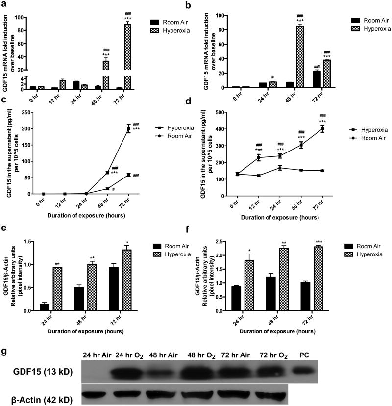
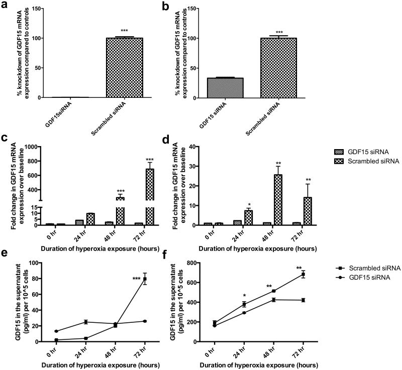
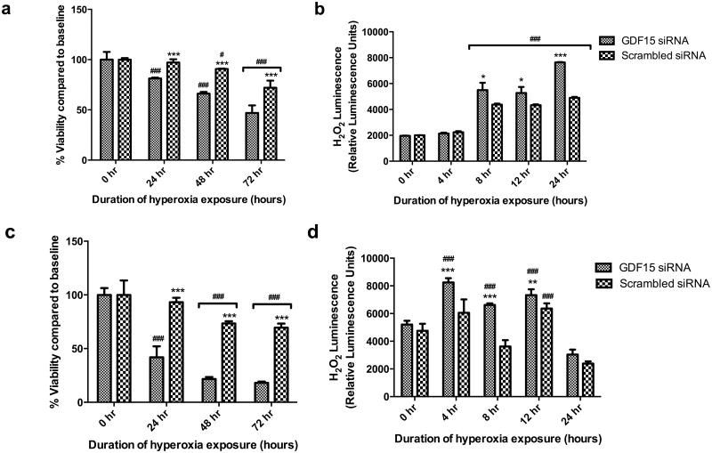
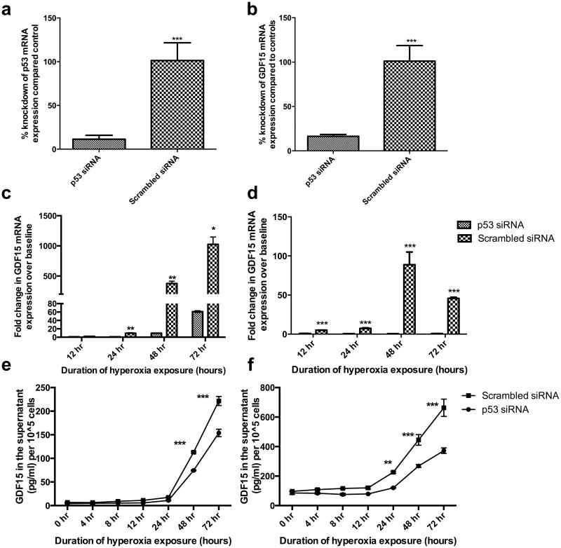
GDF15 (growth and differentiation factor 15) is a secreted cytokine, a direct target of p53 and plays a role in cell proliferation and apoptosis. It is induced by oxidative stress and has anti-apoptotic effects. The role of GDF15 in hyperoxic lung injury is unknown. We tested the hypothesis that GDF15 will be induced in vitro, in a model of pulmonary oxygen toxicity, and will play a critical role in decreasing cell death and oxidative stress. BEAS-2B (human bronchial epithelial cells) and human pulmonary vascular endothelial cells (HPMEC) were exposed to hyperoxia, and expression of GDF15 and effect of GDF15 disruption on cell viability and oxidative stress was determined. Furthermore, we studied the effect of p53 knockdown on GDF15 expression. In vitro, both BEAS-2B and HPMEC cells showed a significant increase in GDF15 expression upon exposure to hyperoxia. After GDF15 knockdown, there was a significant decrease in cell viability and increase in oxidative stress compared to control cells transfected with siRNA with a scrambled sequence. Knockdown of p53 significantly decreased the induction of GDF15 by hyperoxia. In conclusion, we show that GDF15 has a pro-survival and anti-oxidant role in hyperoxia and that p53 plays a key role in its induction.

Keywords: BEAS-2B; Bronchopulmonary dysplasia; HPMEC; Hyperoxia; Pulmonary.


Figures

Figure 1
Figure 1. Induction of GDF15 expression in pulmonary epithelial and endothelial cells in hyperoxia
BEAS-2B (a,c,e,g) and HPMEC (b,d,f) cells exposed to room air (room air-5% CO2) and hyperoxia (95% O2-5% CO2) up to 72 hr. GDF15 expression at the mRNA level (a,b) and at the protein level (ELISA (c,d) and western blot (e-g) was measured. The protein band for GDF15 is the monomer (13kD). β-actin was used as the loading control for the western blot experiments. Values are means ± SEM of 3 independent biological replicates. Significant differences between corresponding room air and hyperoxia exposed cells are indicated by *P <0.05, **P <0.01 and ***P <0.001. Significant differences from 0 hr values are indicated by # P <0.05 and ### P < 0.001.
Figure 2
Figure 2. Knockdown of GDF15 abrogates GDF15 induction in hyperoxia
BEAS-2B (a,c,e) and HPMEC (b,d,f) cells exposed to room air (room air-5% CO2) and 24, 48, or 72 h of hyperoxia (95% O2-5% CO2). GDF15 siRNA or negative control siRNA were transfected into BEAS-2B and HPMEC cells. Expression of GDF-15 was measured at the mRNA (a-d) and protein level (e,f) by ELISA. Values are means ± SEM of 3 independent biological replicates. Significant differences between GDF15 siRNA and negative control siRNA groups are indicated by *P <0.05, **P<0.01 and ***P <0.001.
Figure 3
Figure 3. GDF15 knockdown decreases cell viability and increases oxidative stress in BEAS-2B or HPMEC cells exposed to hyperoxia
BEAS-2B (a,b) and HPMEC (c,d) cells were cultured in room air or hyperoxia were subjected to the MTT assay and ROS-Glo™ luminescent H2O2 assay. Values are means ± SEM of 3 independent biological replicates. Significant differences between GDF15 siRNA and negative control siRNA groups are indicated by *P <0.05, **P<0.01 and ***P <0.001. Significant differences between corresponding 0 hr values are indicated by # P <0.05 and ### P < 0.001.
Figure 4
Figure 4. p53 knockdown decreases GDF15 expression in BEAS-2B or HPMEC cells exposed to hyperoxia
p53 siRNA or negative control siRNA were transfected into BEAS-2B and HPMEC cells. Cells exposed to room air (room air-5% CO2) and up to 72 hr of hyperoxia (95% O2-5% CO2). Figures 4a and 4b show successful p53 knockdown at the mRNA level in BEAS-2B (a) and HPMEC cells (b). Expression of GDF15 was measured at the mRNA (c, d) and protein level (e,f) in BEAS-2B (c, e) and HPMEC (d, f) cells. Values are means ± SEM of 3 independent biological replicates. Significant differences between p53 siRNA and negative control siRNA groups are indicated by *P <0.05, **P<0.01 and ***P <0.001.

Similar articles

Cited by

References

    1. Agarwal MK, Hastak K, Jackson MW, Breit SN, Stark GR, Agarwal ML. Macrophage inhibitory cytokine 1 mediates a p53-dependent protective arrest in S phase in response to starvation for DNA precursors. Proc Natl Acad Sci U S A. 2006;103:16278–16283. doi: 10.1073/pnas.0607210103. - DOI - PMC - PubMed
    1. Böttner M, Suter-Crazzolara C, Schober A, Unsicker K. Expression of a novel member of the TGF-beta superfamily, growth/differentiation factor-15/macrophage-inhibiting cytokine-1 (GDF-15/MIC-1) in adult rat tissues. Cell Tissue Res. 1999;297:103–110. - PubMed
    1. Buczynski BW, Maduekwe ET, O'Reilly MA. The role of hyperoxia in the pathogenesis of experimental BPD. Semin Perinatol. 2013;37:69–78. doi: 10.1053/j.semperi.2013.01.002. - DOI - PMC - PubMed
    1. Clark BJ, Bull TM, Benson AB, Stream AR, Macht M, Gaydos J, Meadows C, Burnham EL, Moss M the ARDS Network Investigators. Growth differentiation factor-15 and prognosis in acute respiratory distress syndrome: a retrospective cohort study. Crit Care. 2013;17:R92. doi: 10.1186/cc12737. - DOI - PMC - PubMed
    1. Han ES, Muller FL, Pérez VI, Qi W, Liang H, Xi L, Fu C, Doyle E, Hickey M, Cornell J, Epstein CJ, Roberts LJ, Van Remmen H, Richardson A. The in vivo gene expression signature of oxidative stress. Physiol Genomics. 2008;34:112–126. doi: 10.1152/physiolgenomics.00239.2007. - DOI - PMC - PubMed

Publication types