

Development of an ultrasonic method to detect cervical remodeling in vivo in full-term pregnant women
- PMID: 26004670
- PMCID: PMC4526398
- DOI: 10.1016/j.ultrasmedbio.2015.04.022
Development of an ultrasonic method to detect cervical remodeling in vivo in full-term pregnant women
Abstract
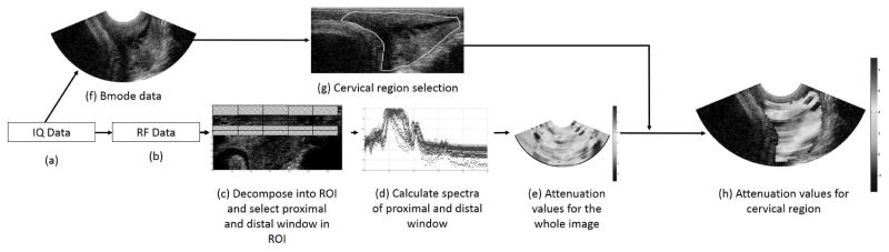
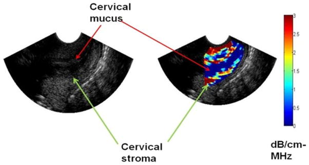
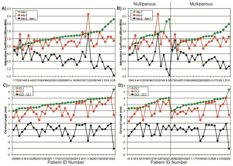
The objective of this study was to determine whether estimates of ultrasonic attenuation could detect changes in the cervix associated with medically induced cervical remodeling. Thirty-six full-term pregnant women underwent two transvaginal ultrasonic examinations separated in time by 12 h to determine cervical attenuation, cervical length and changes thereof. Ultrasonic attenuation and cervical length data were acquired from a zone (Zonare Medical Systems, Mountain View, CA, USA) ultrasound system using a 5-9 MHz endovaginal probe. Cervical attenuation and cervical length significantly decreased in the 12 h between the pre-cervical ripening time point and 12 h later. The mean cervical attenuation was 1.1 ± 0.4 dB/cm-MHz before cervical ripening agents were used and 0.8 ± 0.4 dB/cm-MHz 12 h later (p < 0.0001). The mean cervical length also decreased from 3.1 ± 0.9 cm before the cervical ripening was administered to 2.0 ± 1.1 cm 12 h later (p < 0.0001). Cervical attenuation and cervical length detected changes in cervical remodeling 12 h after cervical ripening administration.
Keywords: Cervical length; Cervical remodeling; Cervical ripening; Ultrasonic attenuation.
Copyright © 2015 World Federation for Ultrasound in Medicine & Biology. Published by Elsevier Inc. All rights reserved.
Figures
Similar articles
-
Beyond Cervical Length: A Pilot Study of Ultrasonic Attenuation for Early Detection of Preterm Birth Risk.Ultrasound Med Biol. 2015 Nov;41(11):3023-9. doi: 10.1016/j.ultrasmedbio.2015.06.014. Epub 2015 Aug 8. Ultrasound Med Biol. 2015. PMID: 26259887 Free PMC article.
-
Ultrasonic attenuation estimation of the pregnant cervix: a preliminary report.Ultrasound Obstet Gynecol. 2010 Aug;36(2):218-25. doi: 10.1002/uog.7643. Ultrasound Obstet Gynecol. 2010. PMID: 20629011 Free PMC article.
-
Role of 3-dimensional power Doppler sonography in differentiating pregnant women with threatened preterm labor from those with an asymptomatic short cervix.J Ultrasound Med. 2014 Apr;33(4):673-9. doi: 10.7863/ultra.33.4.673. J Ultrasound Med. 2014. PMID: 24658947
-
Short cervical length dilemma.Obstet Gynecol Clin North Am. 2015 Jun;42(2):241-54. doi: 10.1016/j.ogc.2015.01.003. Epub 2015 Mar 10. Obstet Gynecol Clin North Am. 2015. PMID: 26002164 Review.
-
The role of sonographic cervical length in labor induction at term.J Clin Ultrasound. 2015 Jan;43(1):7-16. doi: 10.1002/jcu.22229. Epub 2014 Sep 22. J Clin Ultrasound. 2015. PMID: 25243838 Review.
Cited by
-
Cervical Evaluation: From Ancient Medicine to Precision Medicine.Obstet Gynecol. 2017 Jul;130(1):51-63. doi: 10.1097/AOG.0000000000002106. Obstet Gynecol. 2017. PMID: 28594774 Free PMC article.
-
A Phantom-Based Assessment of Repeatability and Reproducibility of Transvaginal Quantitative Ultrasound.IEEE Trans Ultrason Ferroelectr Freq Control. 2019 Sep;66(9):1413-1421. doi: 10.1109/TUFFC.2019.2921925. Epub 2019 Jun 14. IEEE Trans Ultrason Ferroelectr Freq Control. 2019. PMID: 31217100 Free PMC article.
-
Decreased Nutrient Intake Is Associated With Premature Cervical Remodeling.J Obstet Gynecol Neonatal Nurs. 2017 Jan-Feb;46(1):123-134. doi: 10.1016/j.jogn.2016.08.006. Epub 2016 Nov 8. J Obstet Gynecol Neonatal Nurs. 2017. PMID: 27836660 Free PMC article.
-
Heterogeneous microstructural changes of the cervix influence cervical funneling.Acta Biomater. 2022 Mar 1;140:434-445. doi: 10.1016/j.actbio.2021.12.025. Epub 2021 Dec 25. Acta Biomater. 2022. PMID: 34958969 Free PMC article.
-
Repeatability and Reproducibility of the Ultrasonic Attenuation Coefficient and Backscatter Coefficient Measured in the Right Lobe of the Liver in Adults With Known or Suspected Nonalcoholic Fatty Liver Disease.J Ultrasound Med. 2018 Aug;37(8):1913-1927. doi: 10.1002/jum.14537. Epub 2018 Jan 23. J Ultrasound Med. 2018. PMID: 29359454 Free PMC article.
References
-
- Baldwin SL, Yang M, Marutyan KR, Wallace KD, Holland MR, Miller JG. Ultrasonic detection of the anisotropy of protein cross linking in myocardium at diagnostic frequencies. IEEE transactions on ultrasonics, ferroelectrics, and frequency control. 2007;54:1360–9. - PubMed
-
- Bigelow TA, Labyed Y, McFarlin BL, Sen-Gupta E, O’Brien WDJ. Comparison of algorithms for estimating ultrasound attenuation when predicting cervical remodeling in a rat model. Proceedings of the 2011 IEEE International Symposium on Biomedical Imaging. 2011:883–6.
Publication types
MeSH terms
Grants and funding
LinkOut - more resources
Full Text Sources
Other Literature Sources
Medical
Miscellaneous
