

Potential of N-acetylated-para-aminosalicylic acid to accelerate manganese enhancement decline for long-term MEMRI in rodent brain
- PMID: 26004847
- PMCID: PMC4500662
- DOI: 10.1016/j.jneumeth.2015.05.013
Potential of N-acetylated-para-aminosalicylic acid to accelerate manganese enhancement decline for long-term MEMRI in rodent brain
Abstract
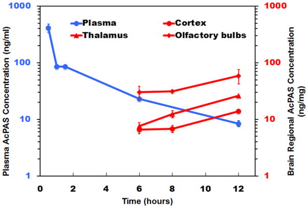
Background: Manganese (Mn(2+))-enhanced MRI (MEMRI) is a valuable imaging tool to study brain structure and function in normal and diseased small animals. The brain retention of Mn(2+) is relatively long with a half-life (t1/2) of 51-74 days causing a slow decline of MRI signal enhancement following Mn(2+) administration. Such slow decline limits using repeated MEMRI to follow the central nervous system longitudinally in weeks or months. This is because residual Mn(2+) from preceding administrations can confound the interpretation of imaging results. We investigated whether the Mn(2+) enhancement decline could be accelerated thus enabling repeated MEMRI, and as a consequence broadens the utility of MEMRI tests.
New methods: We investigated whether N-acetyl-para-aminosalicylic acid (AcPAS), a chelator of Mn(2+), could affect the decline of Mn(2+) induced MRI enhancement in brain.
Results and conclusion: Two-week treatment with AcPAS (200mg/kg/dose×3 daily) accelerated the decline of Mn(2+) induced enhancement in MRI. In the whole brain on average the enhancement declined from 100% to 17% in AcPAS treated mice, while in PBS controls the decline is from 100% to 27%. We posit that AcPAS could enhance MEMRI utility for evaluating brain biology in small animals.
Comparison with existing methods: To the best of our knowledge, no method exists to accelerate the decline of the Mn(2+) induced MRI enhancement for repeated MEMRI tests.
Keywords: Chelation; Manganese enhanced MRI (MEMRI); N-acetylated-para-aminosalicylic acid (AcPAS); Repeated MEMRI.
Copyright © 2015 Elsevier B.V. All rights reserved.
Figures


Similar articles
-
Mn2+ dynamics in manganese-enhanced MRI (MEMRI): Cav1.2 channel-mediated uptake and preferential accumulation in projection terminals.Neuroimage. 2018 Apr 1;169:374-382. doi: 10.1016/j.neuroimage.2017.12.054. Epub 2017 Dec 19. Neuroimage. 2018. PMID: 29277401
-
In Vivo Visualization of Active Polysynaptic Circuits With Longitudinal Manganese-Enhanced MRI (MEMRI).Front Neural Circuits. 2018 May 22;12:42. doi: 10.3389/fncir.2018.00042. eCollection 2018. Front Neural Circuits. 2018. PMID: 29887796 Free PMC article.
-
New method of manganese-enhanced Magnetic Resonance Imaging (MEMRI) for rat brain research.Exp Anim. 2012;61(2):157-64. doi: 10.1538/expanim.61.157. Exp Anim. 2012. PMID: 22531731
-
Longitudinal manganese-enhanced magnetic resonance imaging of neural projections and activity.NMR Biomed. 2022 Jun;35(6):e4675. doi: 10.1002/nbm.4675. Epub 2022 Mar 6. NMR Biomed. 2022. PMID: 35253280 Free PMC article. Review.
-
Applications of manganese-enhanced magnetic resonance imaging (MEMRI) to image brain plasticity in song birds.NMR Biomed. 2004 Dec;17(8):602-12. doi: 10.1002/nbm.936. NMR Biomed. 2004. PMID: 15761949 Review.
Cited by
-
Uptake and retention of manganese contrast agents for PET and MRI in the rodent brain.Contrast Media Mol Imaging. 2016 Sep;11(5):371-380. doi: 10.1002/cmmi.1701. Epub 2016 Jul 11. Contrast Media Mol Imaging. 2016. PMID: 27396476 Free PMC article.
References
-
- Koretsky AP, Silva AC. Manganese-enhanced magnetic resonance imaging (MEMRI) NMR Biomed. 2004 Dec;17(8):527–31. - PubMed
-
- Pautler RG. Biological applications of manganese-enhanced magnetic resonance imaging. Methods Mol Med. 2006;124:365–86. - PubMed
-
- Silva AC, Lee JH, Aoki I, Koretsky AP. Manganese-enhanced magnetic resonance imaging (MEMRI): Methodological and practical considerations. NMR Biomed. 2004 Dec;17(8):532–43. - PubMed
-
- Takeda A. Manganese action in brain function. Brain Res Brain Res Rev. 2003 Jan;41(1):79–87. - PubMed
Publication types
MeSH terms
Substances
Grants and funding
LinkOut - more resources
Full Text Sources
Other Literature Sources
Medical
