

Exposure to HIV-1 Tat in brain impairs sensorimotor gating and activates microglia in limbic and extralimbic brain regions of male mice
- PMID: 26005128
- PMCID: PMC4497922
- DOI: 10.1016/j.bbr.2015.05.021
Exposure to HIV-1 Tat in brain impairs sensorimotor gating and activates microglia in limbic and extralimbic brain regions of male mice
Abstract
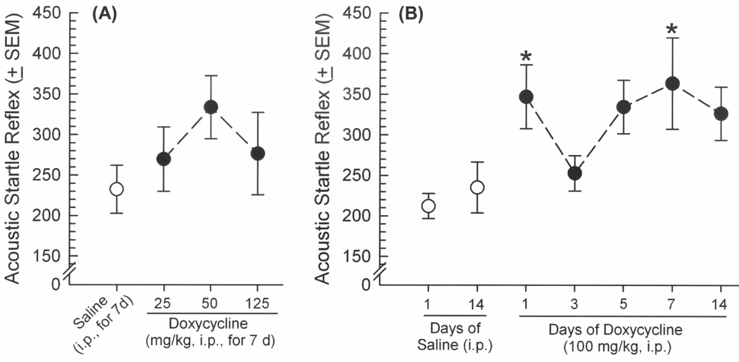
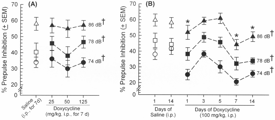
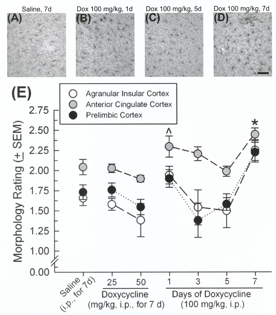
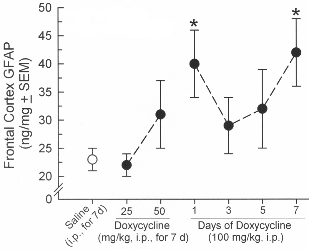
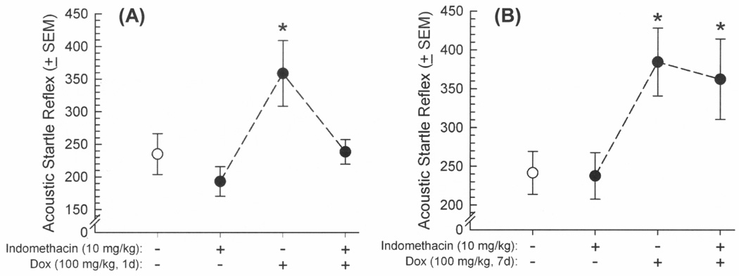
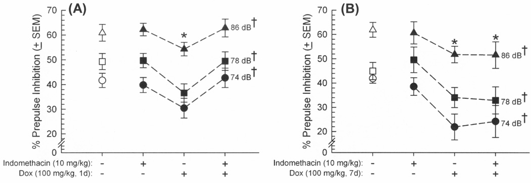
Human immunodeficiency virus (HIV) infection is associated with mood disorders and behavioral disinhibition. Impairments in sensorimotor gating and associated neurocognitive disorders are reported, but the HIV-proteins and mechanisms involved are not known. The regulatory HIV-1 protein, Tat, is neurotoxic and its expression in animal models increases anxiety-like behavior concurrent with neuroinflammation and structural changes in limbic and extra-limbic brain regions. We hypothesized that conditional expression of HIV-1 Tat1-86 in the GT-tg bigenic mouse model would impair sensorimotor gating and increase microglial reactivity in limbic and extralimbic brain regions. Conditional Tat induction via doxycycline (Dox) treatment (0-125 mg/kg, i.p., for 1-14 days) significantly potentiated the acoustic startle reflex (ASR) of GT-tg mice and impaired prepulse inhibition (PPI) of this response in a dose-dependent manner when Dox (100mg/kg) was administered for brief (1 day) or prolonged (daily for 7 days) intervals. A greater proportion of active/reactive Iba1-labeled microglia was seen in the anterior cingulate cortex (ACC), dentate gyrus, and nucleus accumbens core when Tat protein was induced under either brief or prolonged expression conditions. Other subregions of the medial prefrontal cortex, amygdala, hippocampal formation, ventral tegmental area, and ventral pallidum also displayed Tat-induced microglial activation, but only the activation observed in the ACC recapitulated the pattern of ASR and PPI behaviors. Tat exposure also increased frontal cortex GFAP. Pretreatment with indomethacin attenuated the behavioral effects of brief (but not prolonged) Tat-exposure. Overall, exposure to HIV-1 Tat protein induced sensorimotor deficits associated with acute and persistent neuroinflammation in limbic/extralimbic brain regions.
Keywords: Acoustic startle reflex; Glial fibrillary acidic protein (GFAP); Ionized calcium-binding adaptor protein 1; NeuroAIDS; Prepulse inhibition; Trans-activating transcriptor.
Copyright © 2015 Elsevier B.V. All rights reserved.
Figures
Similar articles
-
HIV-1 Tat regulation of dopamine transmission and microglial reactivity is brain region specific.Glia. 2018 Sep;66(9):1915-1928. doi: 10.1002/glia.23447. Epub 2018 May 7. Glia. 2018. PMID: 29733459 Free PMC article.
-
Anxiety-like behavior of mice produced by conditional central expression of the HIV-1 regulatory protein, Tat.Psychopharmacology (Berl). 2014 Jun;231(11):2349-60. doi: 10.1007/s00213-013-3385-1. Epub 2013 Dec 19. Psychopharmacology (Berl). 2014. PMID: 24352568 Free PMC article.
-
Doxycycline-inducible and astrocyte-specific HIV-1 Tat transgenic mice (iTat) as an HIV/neuroAIDS model.J Neurovirol. 2018 Apr;24(2):168-179. doi: 10.1007/s13365-017-0598-9. Epub 2017 Nov 15. J Neurovirol. 2018. PMID: 29143286 Free PMC article. Review.
-
HIV-1 Tat Primes and Activates Microglial NLRP3 Inflammasome-Mediated Neuroinflammation.J Neurosci. 2017 Mar 29;37(13):3599-3609. doi: 10.1523/JNEUROSCI.3045-16.2017. Epub 2017 Mar 7. J Neurosci. 2017. PMID: 28270571 Free PMC article.
-
Multiple actions of the human immunodeficiency virus type-1 Tat protein on microglial cell functions.Neurochem Res. 2004 May;29(5):965-78. doi: 10.1023/b:nere.0000021241.90133.89. Neurochem Res. 2004. PMID: 15139295 Review.
Cited by
-
Immortalization of primary microglia: a new platform to study HIV regulation in the central nervous system.J Neurovirol. 2017 Feb;23(1):47-66. doi: 10.1007/s13365-016-0499-3. Epub 2016 Nov 21. J Neurovirol. 2017. PMID: 27873219 Free PMC article.
-
Conditional Human Immunodeficiency Virus Transactivator of Transcription Protein Expression Induces Depression-like Effects and Oxidative Stress.Biol Psychiatry Cogn Neurosci Neuroimaging. 2017 Oct;2(7):599-609. doi: 10.1016/j.bpsc.2017.04.002. Epub 2017 Apr 20. Biol Psychiatry Cogn Neurosci Neuroimaging. 2017. PMID: 29057370 Free PMC article.
-
Central HIV-1 Tat exposure elevates anxiety and fear conditioned responses of male mice concurrent with altered mu-opioid receptor-mediated G-protein activation and β-arrestin 2 activity in the forebrain.Neurobiol Dis. 2016 Aug;92(Pt B):124-36. doi: 10.1016/j.nbd.2016.01.014. Epub 2016 Feb 1. Neurobiol Dis. 2016. PMID: 26845176 Free PMC article.
-
An Overview of Human Immunodeficiency Virus Type 1-Associated Common Neurological Complications: Does Aging Pose a Challenge?J Alzheimers Dis. 2017;60(s1):S169-S193. doi: 10.3233/JAD-170473. J Alzheimers Dis. 2017. PMID: 28800335 Free PMC article. Review.
-
HIV-1 Tat promotes age-related cognitive, anxiety-like, and antinociceptive impairments in female mice that are moderated by aging and endocrine status.Geroscience. 2021 Feb;43(1):309-327. doi: 10.1007/s11357-020-00268-z. Epub 2020 Sep 17. Geroscience. 2021. PMID: 32940828 Free PMC article.
References
-
- Burdo TH, Katner SN, Taffe MA, Fox HS. Neuroimmunity, drugs of abuse, and neuroAIDS. J Neuroimmune Pharmacol. 2006;1:41–49. - PubMed
-
- Kopnisky KL, Bao J, Lin YW. Neurobiology of HIV, psychiatric and substance abuse comorbidity research: workshop report. Brain Behav Immun. 2007;21:428–441. - PubMed
-
- Copeland KF. Modulation of HIV-1 transcription by cytokines and chemokines. Mini Rev Med Chem. 2005;5:1093–1101. - PubMed
Publication types
MeSH terms
Substances
Grants and funding
LinkOut - more resources
Full Text Sources
Other Literature Sources
Molecular Biology Databases
Research Materials
Miscellaneous
