

PET/MRI assessment of the infarcted mouse heart
- PMID: 26005235
- PMCID: PMC4441008
- DOI: 10.1016/j.nima.2013.08.066
PET/MRI assessment of the infarcted mouse heart
Abstract
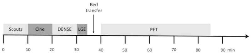
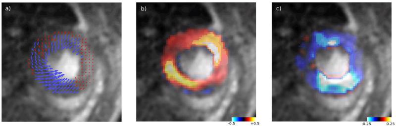
Heart failure originating from myocardial infarction (MI) is a leading cause of death worldwide. Mouse models of ischaemia and reperfusion injury (I/R) are used to study the effects of novel treatment strategies targeting MI, however staging disease and treatment efficacy is a challenge. Damage and recovery can be assessed on the cellular, tissue or whole-organ scale but these are rarely measured in concert. Here, for the first time, we present data showing measures of injury in infarcted mice using complementary techniques for multi-modal characterisation of the heart. We use in vivo magnetic resonance imaging (MRI) to assess heart function with cine-MRI, hindered perfusion with late gadolinium enhancement imaging and muscular function with displacement encoded with stimulated echoes (DENSE) MRI. These measures are followed by positron emission tomography (PET) with 18-F-fluorodeoxyglucose to assess cellular metabolism. We demonstrate a protocol combining each of these measures for the same animal in the same imaging session and compare how the different markers can be used to quantify cardiac recovery on different scales following injury.
Keywords: PET/MRI; mouse; multimodality; myocardial infarction.
Figures


References
-
- Mendis S, et al. Global atlas on cardiovascular disease prevention and control. World Health Organization; 2011.
-
- Schneider JE, Wiesmann F, Lygate CA, Neubauer S. How to perform an accurate assessment of cardiac function in mice using high-resolution magnetic resonance imaging. J Cardiovasc Magn Reson. 2006;8:693–701. doi:10.1080/10976640600723664. - PubMed
-
- Simonetti OP, et al. An improved MR imaging technique for the visualization of myocardial infarction. Radiology. 2001;218:215–223. - PubMed
-
- Buonincontri G, Methner C, Krieg T, Carpenter TA, Sawiak SJ. A fast protocol for infarct quantification in mice. J Magn Reson Imaging. 2013 doi:10.1002/jmri.24001. - PubMed
-
- Gilson WD, Yang Z, French BA, Epstein FH. Measurement of myocardial mechanics inmice before and after infarction using multislice displacement-encoded MRI with 3D motion encoding. Am J Physiol Heart Circ Physiol. 2005;288:H1491–1497. doi:10.1152/ajpheart.00632.2004. - PubMed
Grants and funding
LinkOut - more resources
Full Text Sources
Other Literature Sources