

Selective inhibition of MG-63 osteosarcoma cell proliferation induced by curcumin-loaded self-assembled arginine-rich-RGD nanospheres
- PMID: 26005346
- PMCID: PMC4427601
- DOI: 10.2147/IJN.S78756
Selective inhibition of MG-63 osteosarcoma cell proliferation induced by curcumin-loaded self-assembled arginine-rich-RGD nanospheres
Abstract
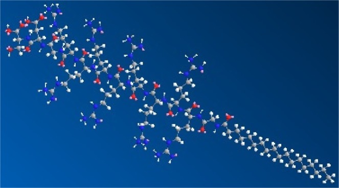
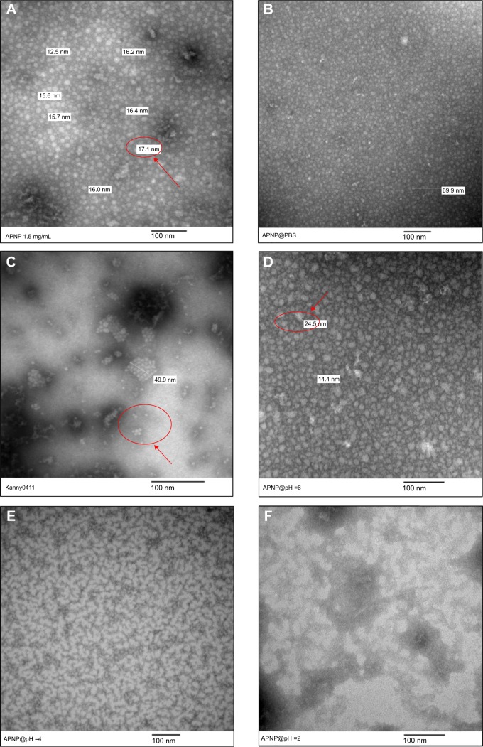
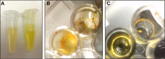
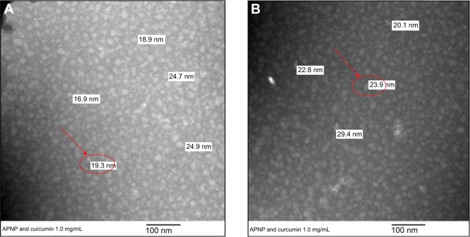
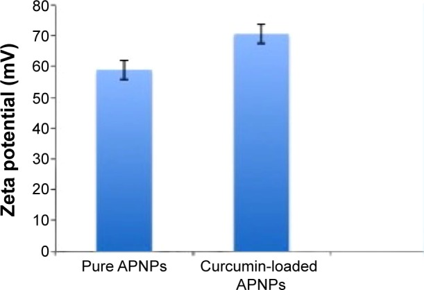
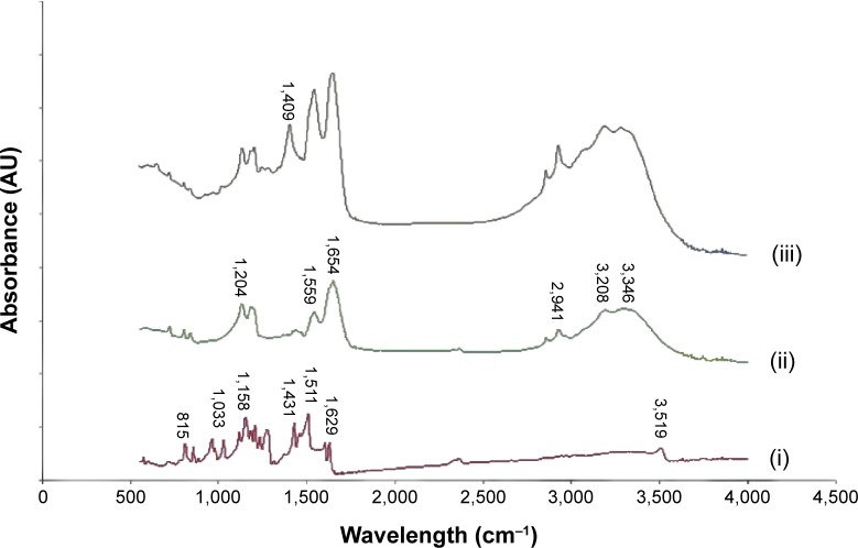
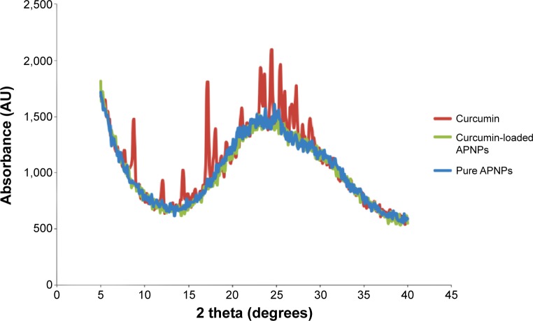
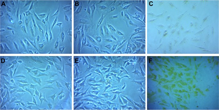
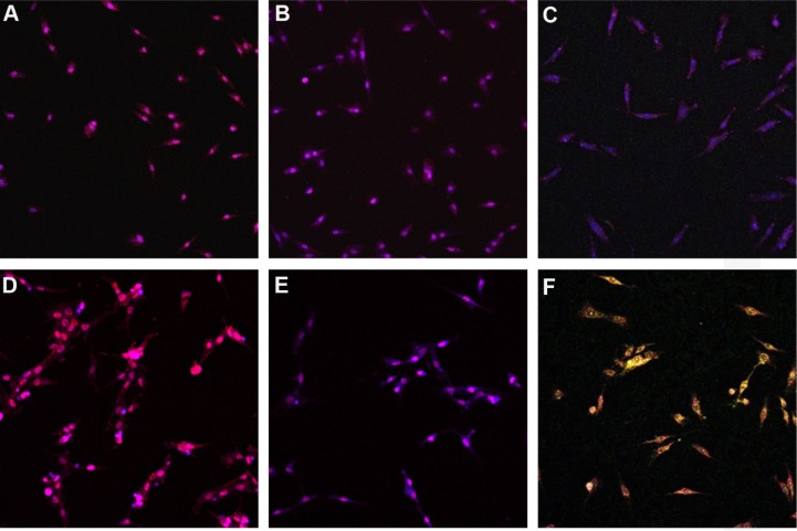
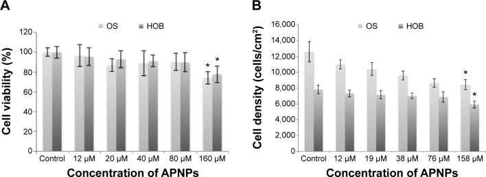
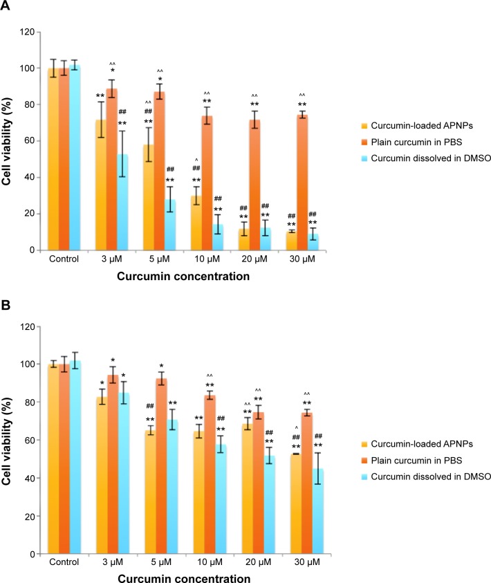
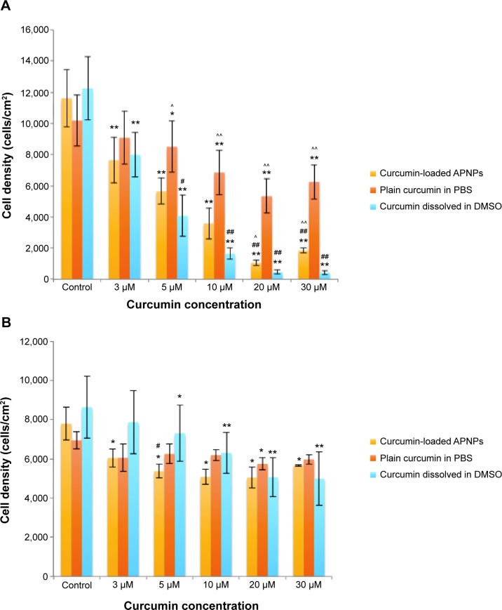
Osteosarcoma is the most frequent primary malignant form of bone cancer, comprising 30% of all bone cancer cases. The objective of this in vitro study was to develop a treatment against osteosarcoma with higher selectivity toward osteosarcoma cells and lower cytotoxicity toward normal healthy osteoblast cells. Curcumin (or diferuloylmethane) has been found to have antioxidant and anticancer effects by multiple cellular pathways. However, it has lower water solubility and a higher degradation rate in alkaline conditions. In this study, the amphiphilic peptide C18GR7RGDS was used as a curcumin carrier in aqueous solution. This peptide contains a hydrophobic aliphatic tail group leading to their self-assembly by hydrophobic interactions, as well as a hydrophilic head group composed of an arginine-rich and an arginine-glycine-aspartic acid structure. Through characterization by transmission electron microscopy, self-assembled structures of spherical amphiphilic nanoparticles (APNPs) with diameters of 10-20 nm in water and phosphate-buffered saline were observed, but this structure dissociated when the pH value was reduced to 4. Using a method of codissolution with acetic acid and dialysis tubing, the solubility of curcumin was enhanced and a homogeneous solution was formed in the presence of APNPs. Successful encapsulation of curcumin in APNPs was then confirmed by Fourier transform infrared and X-ray diffraction analyses. The cytotoxicity and cellular uptake of the APNP/curcumin complexes on both osteosarcoma and normal osteoblast cell lines were also evaluated by methyl-thiazolyl-tetrazolium assays and confocal fluorescence microscopy. The results showed that the curcumin-loaded APNPs had significant selective cytotoxicity against MG-63 osteosarcoma cells when compared with normal osteoblasts. We have demonstrated for the first time that APNPs can encapsulate hydrophobic curcumin in their hydrophobic cores, and curcumin-loaded APNPs could be an innovative treatment for the selective inhibition of osteosarcoma cells.
Keywords: arginine-glycine-aspartic acid; arginine-rich; curcumin; osteosarcoma; selective inhibition; self-assembly.
Figures
Similar articles
-
Curcumin and its Analogs and Carriers: Potential Therapeutic Strategies for Human Osteosarcoma.Int J Biol Sci. 2023 Feb 13;19(4):1241-1265. doi: 10.7150/ijbs.80590. eCollection 2023. Int J Biol Sci. 2023. PMID: 36923933 Free PMC article. Review.
-
Design of polyaspartic acid peptide-poly (ethylene glycol)-poly (ε-caprolactone) nanoparticles as a carrier of hydrophobic drugs targeting cancer metastasized to bone.Int J Nanomedicine. 2017 May 8;12:3561-3575. doi: 10.2147/IJN.S133787. eCollection 2017. Int J Nanomedicine. 2017. PMID: 28507436 Free PMC article.
-
Formulation, characterization and evaluation of curcumin-loaded PLGA nanospheres for cancer therapy.Anticancer Res. 2009 Oct;29(10):3867-75. Anticancer Res. 2009. PMID: 19846921
-
Dual targeting curcumin loaded alendronate-hyaluronan- octadecanoic acid micelles for improving osteosarcoma therapy.Int J Nanomedicine. 2019 Aug 9;14:6425-6437. doi: 10.2147/IJN.S211981. eCollection 2019. Int J Nanomedicine. 2019. PMID: 31496695 Free PMC article.
-
Curcumin in human osteosarcoma: From analogs to carriers.Drug Discov Today. 2023 Feb;28(2):103437. doi: 10.1016/j.drudis.2022.103437. Epub 2022 Nov 11. Drug Discov Today. 2023. PMID: 36372327 Review.
Cited by
-
Production of Curcumin-Loaded Silk Fibroin Nanoparticles for Cancer Therapy.Nanomaterials (Basel). 2018 Feb 24;8(2):126. doi: 10.3390/nano8020126. Nanomaterials (Basel). 2018. PMID: 29495296 Free PMC article.
-
Antioxidant and Cytotoxic Effects of the Methanolic Extract of Eichhornia crassipes Petioles Upon Mg-63 Cell Lines: An In Vitro Study.Cureus. 2023 May 2;15(5):e38425. doi: 10.7759/cureus.38425. eCollection 2023 May. Cureus. 2023. PMID: 37273397 Free PMC article.
-
Curcumin-Loaded Nanoparticles Impair the Pro-Tumor Activity of Acid-Stressed MSC in an In Vitro Model of Osteosarcoma.Int J Mol Sci. 2021 May 28;22(11):5760. doi: 10.3390/ijms22115760. Int J Mol Sci. 2021. PMID: 34071200 Free PMC article.
-
Curcumin and its Analogs and Carriers: Potential Therapeutic Strategies for Human Osteosarcoma.Int J Biol Sci. 2023 Feb 13;19(4):1241-1265. doi: 10.7150/ijbs.80590. eCollection 2023. Int J Biol Sci. 2023. PMID: 36923933 Free PMC article. Review.
-
Synthesis and study of cell-penetrating peptide-modified gold nanoparticles.Int J Nanomedicine. 2018 Oct 9;13:6199-6205. doi: 10.2147/IJN.S168720. eCollection 2018. Int J Nanomedicine. 2018. PMID: 30349244 Free PMC article.
References
-
- He JP, Hao Y, Wang XL, et al. Review of the molecular pathogenesis of osteosarcoma. Asian Pac J Cancer Prev. 2014;15(15):5967–5976. - PubMed
-
- Cotterill SJ, Wright CM, Pearce MS, Craft AW. Stature of young people with malignant bone tumors. Pediatr Blood Cancer. 2004;42(1):59–63. - PubMed
-
- Ottaviani G, Jaffe N. The Etiology of Osteosarcoma. In: Jaffe N, Bruland ØS, Bielack SS, editors. Pediatric and Adolescent Osteosarcoma. Houston, TX, USA: Springer; 2009. pp. 15–32. - PubMed
Publication types
MeSH terms
Substances
LinkOut - more resources
Full Text Sources
Other Literature Sources
Medical
