. 2015 May 7:8:29-37.
doi: 10.2147/JAA.S75261. eCollection 2015.

Activated protein C modulates the proinflammatory activity of dendritic cells

Affiliations

Activated protein C modulates the proinflammatory activity of dendritic cells

Takahiro Matsumoto et al. J Asthma Allergy. .

Abstract

Background: Previous studies have demonstrated the beneficial activity of activated protein C in allergic diseases including bronchial asthma and rhinitis. However, the exact mechanism of action of activated protein C in allergies is unclear. In this study, we hypothesized that pharmacological doses of activated protein C can modulate allergic inflammation by inhibiting dendritic cells.

Materials and methods: Dendritic cells were prepared using murine bone marrow progenitor cells and human peripheral monocytes. Bronchial asthma was induced in mice that received intratracheal instillation of ovalbumin-pulsed dendritic cells.

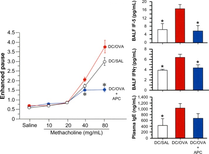
Results: Activated protein C significantly increased the differentiation of tolerogenic plasmacytoid dendritic cells and the secretion of type I interferons, but it significantly reduced lipopolysaccharide-mediated maturation and the secretion of inflammatory cytokines in myeloid dendritic cells. Activated protein C also inhibited maturation and the secretion of inflammatory cytokines in monocyte-derived dendritic cells. Activated protein C-treated dendritic cells were less effective when differentiating naïve CD4 T-cells from Th1 or Th2 cells, and the cellular effect of activated protein C was mediated by its receptors. Mice that received adoptive transfer of activated protein C-treated ovalbumin-pulsed dendritic cells had significantly less airway hyperresponsiveness, significantly decreased lung concentrations of Th1 and Th2 cytokines, and less plasma concentration of immunoglobulin E when compared to control mice.

Conclusion: These results suggest that dendritic cells mediate the immunosuppressive effect of activated protein C during allergic inflammation.

Keywords: allergy; coagulation; dendritic cells; protein C pathway.


Figures

Figure 1
Figure 1
APC promotes the expansion of plasmacytoid DCs. Notes: Bone marrow cells were prepared from C57BL/6 mice and cultured in the presence of Flt3L, and then 20 μg/mL of APC was added to the culture on day 4 and further cultured for 3 days. The proportion of pDCs (CD45R+, CD11b), cDCs (CD45R, CD11b+), and the ratio of cDCs to pDCs (A), the secretion of cytokines from DCs (B), and the RT-PCR analysis of IFNα and IFNβ (C) are described. The data are expressed as the mean ± standard error of the mean. The figure shows the representative result from one of three independent experiments. *P<0.05 compared to the untreated group. Abbreviations: APC, activated protein C; IL, interleukin; DCs, dendritic cells; cDC, conventional dendritic cells; pDCs, plasmacytoid dendritic cells; GAPDH, glyceraldehyde 3-phosphate dehydrogenase; IFN, interferon; RT-PCR, reverse transcription polymerase chain reaction.
Figure 2
Figure 2
APC inhibits maturation of bone marrow-derived DCs. Notes: Bone marrow cells were prepared from C57BL/6 mice, cultured in the presence of Flt3L, and then APC was added to the culture on day 6 and stimulated with LPS for 24 hours. The expression of MHC class II, CD86, and CD80 on CD11c+ DCs in the presence of 40 μg/mL of APC (A) and the mean fluorescent intensity were measured (B). The effects of different doses of APC on maturation markers and secretion of cytokines from DCs (C) are also shown. Data are expressed as the mean ± standard error of the mean (n=3). The figure shows the representative result from one of three independent experiments. *P<0.05 compared to the untreated group. Abbreviations: APC, activated protein C; MHC, major histocompatibility complex; MFI, mean fluorescence intensity; IL, interleukin; DCs, dendritic cells; LPS, lipopolysaccharide; n, number.
Figure 3
Figure 3
APC inhibits the maturation of human monocyte-derived DCs. Notes: Monocytes were prepared from human peripheral blood and cultured in the presence of recombinant GM-CSF and IL-4, and then APC (40 μg/mL) was added to the culture on day 6 and stimulated with LPS for 24 hours. The expression of HLA-DR, CD86, and CD80 on CD11c+ DCs in the presence or absence of APC (A), as well as the secretion of TNFα from DCs (B) are shown. Data are expressed as the mean ± standard error of the mean. The figure shows the representative result from one of three independent experiments. *P<0.05 compared to the untreated group. Abbreviations: Max, maximum; APC, activated protein C; MFI, mean fluorescence intensity; GAPDH, glyceraldehyde 3-phosphate dehydrogenase; TNFα, tumor necrosis factor alpha; DCs, dendritic cells; GM-CSF, granulocyte macrophage colony-stimulating factor; IL, interleukin; LPS, lipopolysaccharide.
Figure 4
Figure 4
APC-treated bone marrow-derived DCs are less effective to prime naïve CD4 T-cells. Notes: Bone marrow cells were prepared and cultured in the presence of recombinant human Flt3L; then, doses of APC were added. Naïve CD4 T-cells were prepared and then stimulated with CD11c+ DCs either in the Th1-skewing condition (A) or the Th2-skewing condition (B). The relative proportion of IFNγ+ CD4 T-cells in the Th1-skewing condition, and IL-4+ and IL-10+ CD4 T-cells in Th2-skewing condition are shown. Data are expressed as the mean ± standard error of the mean. The figure shows the representative result from one of the three independent experiments. *P<0.05 compared to the untreated group. Abbreviations: IFN, interferon; APC, activated protein C; IL, interleukin; DCs, dendritic cells.
Figure 5
Figure 5
Mediation of APC receptors in the bone marrow-derived DC-mediated priming of T-cells. Notes: Bone marrow-derived DCs were prepared and cultured in the presence of recombinant Flt3L for 6 days and stimulated with LPS for 24 hours. Total RNA was prepared from immature (LPS-untreated) and mature (LPS-treated) DCs and analyzed for the expression of EPCR, PAR-1, PAR-2, PAR-3, and PAR-4 by RT-PCR (A). Expressions of EPCR, PAR-1, and CD11b on the cell surface of DCs were analyzed by flow cytometry (A). For T-cell priming assays, naïve CD4 T-cells were prepared and then cultured with CD11c+ DCs. The relative proportion of IFNγ+ CD4 T-cells and IFNγ secretion (B), as well as RNA expression of IFNγ, IL-4, and IL-10 (C) are shown. Data are expressed as the mean ± standard error of the mean (n=3). The figure shows the representative result from one of two independent experiments. *P<0.05 compared to the untreated group. **P<0.01 compared to the group treated with APC alone. Abbreviations: EPCR, endothelial protein C receptor; PAR, protease-activated receptor; DCs, dendritic cells; Max, maximum; IFN, interferon; GAPDH, glyceraldehyde 3-phosphate dehydrogenase; APC, activated protein C; IL, interleukin; LPS, lipopolysaccharide; RT-PCR, reverse transcription polymerase chain reaction; n, number.
Figure 6
Figure 6
APC inhibits DC activation in vivo. Notes: Bone marrow-derived DCs were pulsed with OVA, treated with APC, instilled into the trachea of mice, and then the mice were challenged with the antigen. Hyperresponsiveness was measured by enhanced pause and the concentrations of IL-5, IFNγ, and IgE were measured by ELISA. Data are expressed as the mean ± standard error of the mean (n=3). The figure shows the representative result from one of two independent experiments. *P<0.05 compared to the DC/OVA group. Abbreviations: DC, dendritic cell; OVA, ovalbumin; SAL, saline; APC, activated protein C; BALF, bronchoalveolar lavage fluid; IL, interleukin; IFN, interferon; Ig, immunoglobulin; ELISA, enzyme-linked immunosorbent assay; n, number.

References

    1. Boltjes A, van Wijk F. Human dendritic cell functional specialization in steady-state and inflammation. Front Immunol. 2014;5:131. - PMC - PubMed
    1. Kadowaki N. The divergence and interplay between pDC and mDC in humans. Front Biosci (Landmark Ed) 2009;14:808–817. - PubMed
    1. Randolph GJ, Ochando J, Partida-Sánchez S. Migration of dendritic cell subsets and their precursors. Annu Rev Immunol. 2008;26:293–316. - PubMed
    1. Suzuki K, Gabazza EC, Hayashi T, Kamada H, Adachi Y, Taguchi O. Protective role of activated protein C in lung and airway remodeling. Crit Care Med. 2004;32(5 Suppl):S262–S265. - PubMed
    1. Castellino FJ, Ploplis VA. The protein C pathway and pathologic processes. J Thromb Haemost. 2009;7(Suppl 1):140–145. - PMC - PubMed