Review
. 2015 May 8:9:74.
doi: 10.3389/fnsys.2015.00074. eCollection 2015.

Constructing the suprachiasmatic nucleus: a watchmaker's perspective on the central clockworks

Affiliations
Review

Constructing the suprachiasmatic nucleus: a watchmaker's perspective on the central clockworks

Joseph L Bedont et al. Front Syst Neurosci. .

Abstract

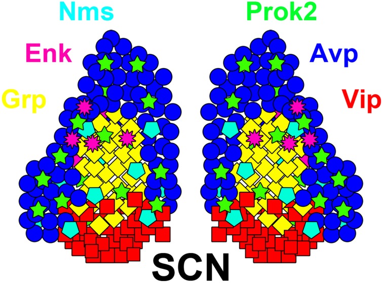
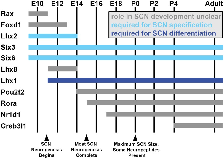
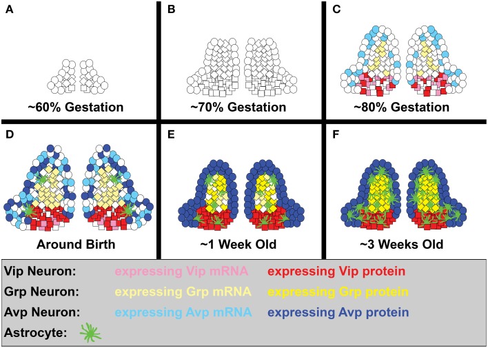
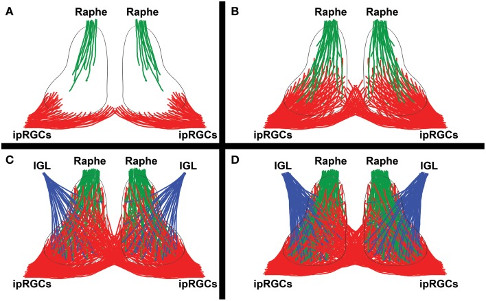
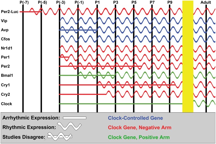
The circadian system constrains an organism's palette of behaviors to portions of the solar day appropriate to its ecological niche. The central light-entrained clock in the suprachiasmatic nucleus (SCN) of the mammalian circadian system has evolved a complex network of interdependent signaling mechanisms linking multiple distinct oscillators to serve this crucial function. However, studies of the mechanisms controlling SCN development have greatly lagged behind our understanding of its physiological functions. We review advances in the understanding of adult SCN function, what has been described about SCN development to date, and the potential of both current and future studies of SCN development to yield important insights into master clock function, dysfunction, and evolution.

Keywords: axon pathfinding; cell fate; chronotype; circadian; clock genes; developmental neuroscience; suprachiasmatic; transcription factors.


Figures

Figure 1
Figure 1
Neuron diversity in the adult SCN. A diagram of the adult SCN, showing a subset of its many neuropeptidergic populations. Neuropeptide colors are indicated by their names, positioned around the SCN. Note that many of these neuropeptides cross or exist outside of the classical core and shell SCN subdomains. Also note that many of these neurotransmitters are at least partially co-expressed, such as in Vip/Grp neurons, though the various probable combinations are not shown here for simplicity. Adapted from Bedont et al. (2014) Graphical Abstract.
Figure 2
Figure 2
SCN transcription factor expression during development. Estimated ages of expression for a subset of hypothalamus- and SCN-enriched transcription factors in the developing mouse SCN between embryonic day (E) 10 and adulthood, selected based on interesting expression patterns and/or known developmental functions (indicated by bar color). Note that many general hypothalamic transcription factors (expressed prior to E10) are downregulated as the transcriptional network controlling SCN development ramps up. Not shown in this figure, other transcription factors expressed throughout the SCN during its development in turn become progressively compartmentalized to specific subdomains as the SCN matures (ex: Lhx1, Rorα).
Figure 3
Figure 3
SCN neurogenesis and differentiation. A simplified diagram showing the development of Vip, Grp, and Avp neurons, as well as astrocytes, in the rodent SCN. While domain boundaries are depicted as being relatively static here to aid in illustrating our points, note that they are actually quite dynamic over the course of development. (A) Early SCN neurogenesis begins at ~60% of gestation (hamster E9.5, mouse E12, and rat E13.5). (B) By ~70% of gestation (hamster E11, mouse E13.5, rat E15), most ventrolateral neurons have already been produced, while dorsomedial neurogenesis is still in full swing. (C) The final major burst of SCN neurogenesis (including a number of ventrolateral neurons) occurs at ~80% of gestation (hamster E12.5, mouse E15, rat E17). Shortly thereafter, many SCN neuropeptide transcripts, but only Vip protein, are detectable (hamster E13, mouse E18-P0). (D) Shortly before and after birth, the first SCN astroglia appear (hamster E15, rat E20), Avp protein expression becomes detectable (P1), and SCN neuron number peaks (hamster P2). (E) By roughly a week into life, a major jump in astrocyte number has occurred (rat P3-P4), cell death brings neuron numbers down to adult levels (hamster P5, mouse P7, rat P6), and Grp protein expression is detectable (hamster P8). (F) By the end of the second major jump in astrocyte number (rat P20-25), the SCN is largely mature. At this stage, the fate of its neurons and the subdomain boundaries they form are relatively stable, though they may remain somewhat malleable under circadian challenge from the environment.
Figure 4
Figure 4
Innervation by major SCN afferents. A simplified diagram showing the development of major SCN afferents from the intrinsically photosensitive retinal ganglion cells (ipRGCs) of the retina, raphe nuclei, and intergeniculate leaflet (IGL). (A) ipRGCs first begin to innervate the vlSCN ipsilaterally at birth (mouse/rat P0-P1) or shortly thereafter (hamster P3). The raphe nuclei first innervate the SCN simultaneously (mouse P0, hamster P3). (B) A few days later, ipRGC innervation of the SCN becomes noticeably denser and broader, with the first contralateral projections appearing in the ventromedial SCN (mouse/rat P4), as raphe innervation also builds. (C) By roughly a week into postnatal SCN development, raphe innervation has become fairly dense (mouse P5, hamster P6), ipRGC contralateral and ipsilateral ipRGC innervation domains are commingled (mouse P7), and the earliest Npy signal from the IGL is detectable (hamster P7, rat P10). Many of the IGL and raphe projections converge, a trend that persists through adulthood. (D) In the following few weeks, more-or-less adult SCN innervation by the raphe (mouse P10), ipRGC (mouse P14), and IGL (rat P20) projections become apparent.
Figure 5
Figure 5
Ontogeny of the SCN clockworks. A simplified diagram showing the approximate age at which expression and cycling of various cellular clock and clock-controlled genes are known to begin in the SCN. Data from all mouse and rat studies we were able to locate is included in the figure; our P(-x) terminology is used for prenatal time-points to roughly account for the difference in average gestation time in mouse (P0 at ~20 days) and rat (P0 at ~22 days). In vivo studies were surprisingly consistent despite differences in organism and detection methodology among studies, arriving at estimated ages of rhythm ontogeny within a few day window in most cases. Notably late outliers include Per2 (Shimomura et al., 2001) and Cry1 (Ansari et al., 2009). New studies observing Per2-Luc luminescence in slice have arrived at earlier estimates of rhythm induction than in vivo studies of Per2; however, the reason for this is unclear (see text). Finally, note that only induction of expression and rhythmic expression are accurately recapitulated in this figure; relative phase and amplitude of the peaks is incidental.

References

    1. Abe M., Herzog E. D., Yamazaki S., Straume M., Tei H., Sakaki Y., et al. (2002). Circadian rhythms in isolated brain regions. J. Neurosci. 22, 350–356. - PMC - PubMed
    1. Abizaid A., Mezei G., Sotonyi P., Horvath T. L. (2004). Sex differences in adult suprachiasmatic nucleus neurons emerging late prenatally in rats. Eur. J. Neurosci. 19, 2488–2496. 10.1111/j.0953-816X.2004.03359.x - DOI - PubMed
    1. Abrahamson E. E., Moore R. Y. (2001). Suprachiasmatic nucleus in the mouse: retinal innervation, intrinsic organization and efferent projections. Brain Res. 916, 172–191. - PubMed
    1. Abrahamson E. E., Moore R. Y. (2006). Lesions of suprachiasmatic nucleus efferents selectively affect rest-activity rhythm. Mol. Cell. Endocrinol. 252, 46–56. 10.1016/j.mce.2006.03.036 - DOI - PubMed
    1. Ahern T. H., Krug S., Carr A. V., Murray E. K., Fitzpatrick E., Bengston L., et al. (2013). Cell death atlas of the postnatal mouse ventral forebrain and hypothalamus: effects of age and sex. J. Comp. Neurol. 521, 2551–2569. 10.1002/cne.23298 - DOI - PMC - PubMed

LinkOut - more resources