. 2015 Feb;6(1):290.
doi: 10.4172/2155-9899.1000290.

Involvement of Heparanase in Empyema: Implication for Novel Therapeutic Approaches

Affiliations

Involvement of Heparanase in Empyema: Implication for Novel Therapeutic Approaches

Moshe Lapidot et al. J Clin Cell Immunol. 2015 Feb.

Abstract

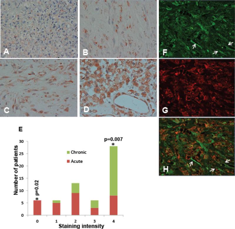
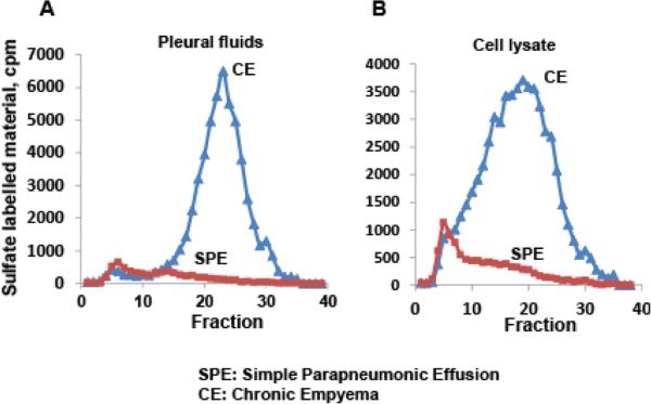
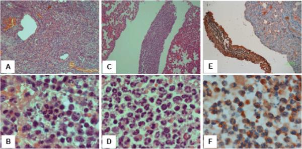
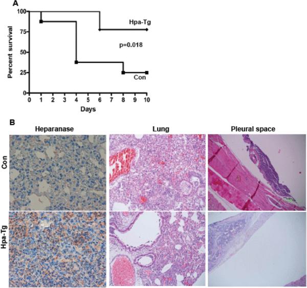
Pleural empyema is an inflammatory condition that progresses from acute to chronic, life-threatening, phase. The incidence of empyema has been increasing both in children and adults worldwide in the past decades, mainly in healthy young adults and in older patients. Despite continued advances in the management of this condition, morbidity and mortality have essentially remained static over the past decade. Better understanding of the disease and the development of new therapeutic approaches are thus critically needed. Heparanase is an endoglucuronidase that cleaves heparan sulfate chains of proteoglycans. These macromolecules are most abounded in the sub-endothelial and sub-epithelial basement membranes and their cleavage by heparanase leads to disassembly of the extracellular matrix that becomes more susceptible to extravasation and dissemination of metastatic and immune cells. Here, we provide evidence that heparanase expression and activity are markedly increased in empyema and pleural fluids, associating with disease progression. Similarly, heparanase expression is increased in a mouse model of empyema initiated by intranasal inoculation of S. pneumonia. Applying this model we show that transgenic mice over expressing heparanase are more resistant to the infection and survive longer.

Keywords: Chronic inflammation; Empyema; Heparanase; Transgenic mice.


Figures

Figure 1
Figure 1
Heparanase elevation in pleural empyema. A-D: Immunostaining. Specimens from 46 empyema patients were subjected to immunostaining applying anti-heparanase antibody. Shown are representative photomicrographs of biopsies exhibiting very weak (+1; A), weak (+2; B), moderate (+3; C) and strong (+4; D) staining of heparanase. Very weak staining (+1) was most often observed in acute empyema while chronic empyema is associated with higher levels of heparanase (B-D). This association is shown graphically in (E). Original magnification: A-D x40. F-H: Immunofluorescent staining. Specimens of chronic empyema were subjected to immunofluorescent staining applying anti heparanase (F) and anti CD163 (human macrophage marker; G) antibodies. Merged image is shown in (H). (arrows). Shown are representative photomicrographs; Original magnification: x40. Note that heparanase staining is cytoplasmic whereas CD163 labels a membrane determinant, and that heparanase also labels CD163-negative cells that are suspected (by morphology) to be endothelial cells lining lumen-containing structures (arrows).
Figure 2
Figure 2
Heparanase activity. Fluids were freshly collected from chronic empyema (CE) patients or patients with simple paraneumonic effusion (SPE); Fluids were centrifuged (300 g, 10 min), and the supernatants and cell pellets were recovered and evaluated for heparanase enzymatic activity as described under ‘Materials and Methods’. Shown are typical heparanase activities (i.e., release of HS degradation fragments from sulfate labeled ECM) in the clear fluids (A) and cell lysates (B).
Figure 3
Figure 3
Mouse model of empyema. Mice were inoculated intranasally with 2 × 10⁸ CFU of S. pneumonia (strain D39). Control mice were inoculated with equal volume of saline. Mice were sacrificed 3 days after inoculation and pleural fluid was collected and cleared by centrifugation; Lung tissue was harvested, fixed, embedded in paraffin and subjected to pathological evaluation and immunohistochemical analysis. Shown are representative H&E staining of the inflamed lung (A, B), and pleural space (C, D). Inflammatory cells in the pleural space are stained positive for heparanase (E, F). Original magnification: A, C, E x10; B, D, F x100.
Figure 4
Figure 4
Heparanase over-expressing transgenic mice exhibit prolonged survival following induction of empyema. Control Balb/C (Con) and heparanase transgenic (Hpa-Tg) mice were inoculated with 2 × 10⁸ CFU of S. pneumonia bacteria and survival of the mice was recorded (A). Lung tissue was collected from surviving mice and postmortem, fixed in formalin and embedded in paraffin. Shown are representative H&E staining of lung specimens (B, middle panels) and pleural space (B, right panels). Specimens were also subjected to immunostaining applying anti-heparanase antibody (left panels), depicting over expression of heparanase in lung tissue of Hpa-Tg mice. Note the lack of inflammation in the pleural space and prolonged survival of Hpa-Tg vs. control wild type mice. Original magnification: left panels x40; middle and right panels x10.

Similar articles

Cited by

References

    1. Alfageme I, Muñoz F, Peña N, Umbría S. Empyema of the thorax in adults. Etiology, microbiologic findings, and management. Chest. 1993;103:839–843. - PubMed
    1. Kwon YS. Pleural infection and empyema. Tuberculosis and respiratory diseases. 2014;76:160–162. - PMC - PubMed
    1. Kroegel C, Antony VB. Immunobiology of pleural inflammation: potential implications for pathogenesis, diagnosis and therapy. Eur Respir J. 1997;10:2411–2418. - PubMed
    1. Aleman C, Alegre J, Monasterio J, Segura RM, Armadans L, et al. Association between inflammatory mediators and the fibrinolysis system in infectious pleural effusions. Clinical Science. 2003;105:601–607. - PubMed
    1. Mutsaers SE, Prele CM, Brody AR, Idell S. Pathogenesis of pleural fibrosis. Respirology. 2004;9:428–440. - PubMed

LinkOut - more resources