. 2015 Jun;17(6):736-48.
doi: 10.1038/ncb3174. Epub 2015 May 25.

Haemocytes control stem cell activity in the Drosophila intestine

Affiliations

Haemocytes control stem cell activity in the Drosophila intestine

Arshad Ayyaz et al. Nat Cell Biol. 2015 Jun.

Abstract

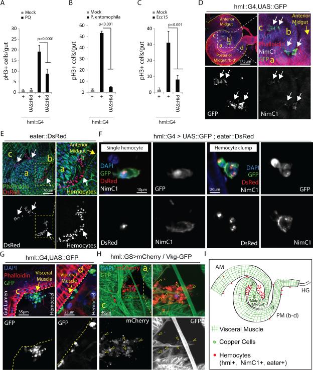
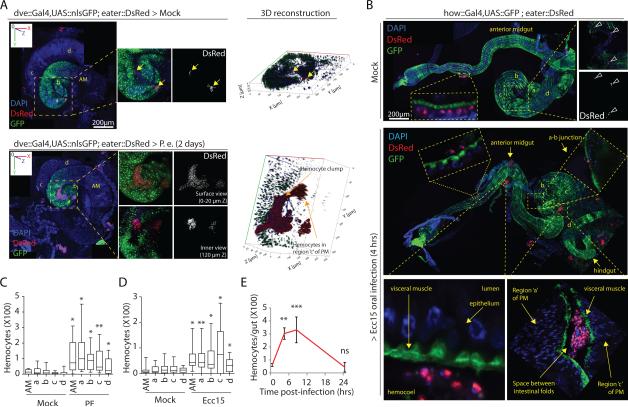
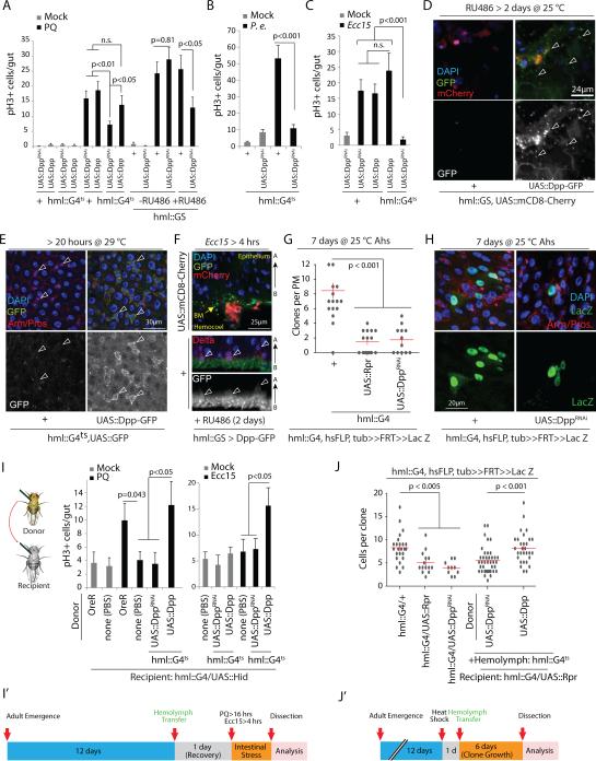
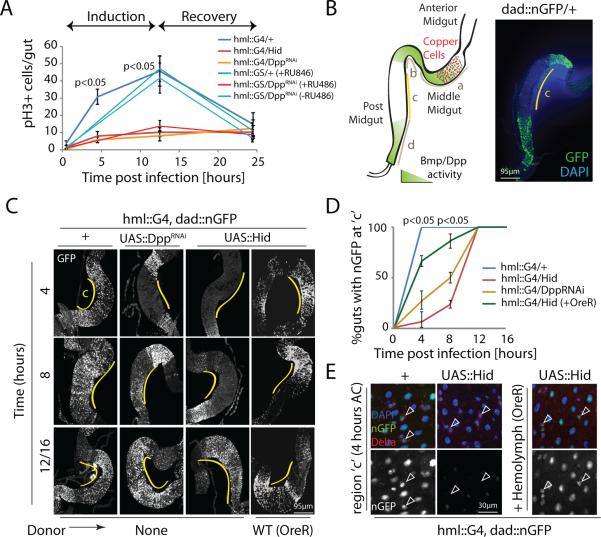
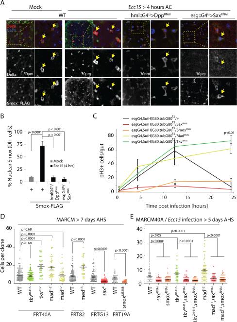
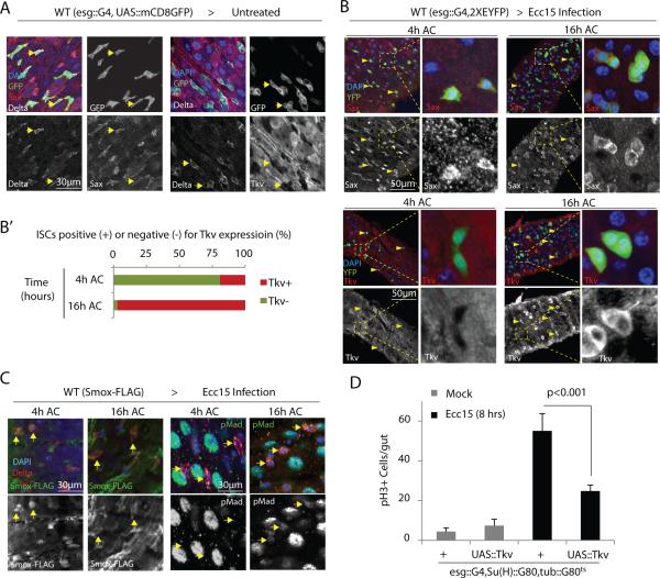
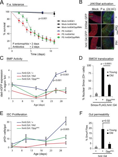
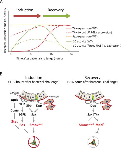
Coordination of stem cell activity with inflammatory responses is critical for regeneration and homeostasis of barrier epithelia. The temporal sequence of cell interactions during injury-induced regeneration is only beginning to be understood. Here we show that intestinal stem cells (ISCs) are regulated by macrophage-like haemocytes during the early phase of regenerative responses of the Drosophila intestinal epithelium. On tissue damage, haemocytes are recruited to the intestine and secrete the BMP homologue DPP, inducing ISC proliferation by activating the type I receptor Saxophone and the Smad homologue SMOX. Activated ISCs then switch their response to DPP by inducing expression of Thickveins, a second type I receptor that has previously been shown to re-establish ISC quiescence by activating MAD. The interaction between haemocytes and ISCs promotes infection resistance, but also contributes to the development of intestinal dysplasia in ageing flies. We propose that similar interactions influence pathologies such as inflammatory bowel disease and colorectal cancer in humans.


Figures

Figure 1
Figure 1. Gut-associated hemocytes are required for ISC proliferation
(A-C) Mitotic activity of ISCs (identified by phospho-HistoneH3-positive cells) in intestines of 3 day-old hemoless flies exposed to Paraquat: PQ (A), Pseudomonas entomophila (B) or Ecc15 (C) is compared with wild-type controls. (D,E) Hemocytes associated with the fly gut under normal rearing conditions can be detected using multiple hemocyte markers: HmlΔ::Gal4 driver and NimC1 (D; white arrows) or eater::DsRed reporter (E; white arrows), while arbitrary midgut regions are identified morphologically using Phalloidin staining that marks Actin (D,E; also see Supplementary Fig.2G). (F) Hemocytes attach fly intestine as single cells or clumps of irregular shape and express all three hemocyte markers: hml (Hemolectin), eater and NimC1 (Nimrod). Note that hemocyte reporter nuclear eaterDsRed provides efficient quantification for actual number of hemocytes per clump. (G) Single hemocytes or clumps (white arrows) attach midgut at the outer surface of visceral muscle (marked by Phalloidin staining; yellow arrows); where c and d: posterior midgut regions c and d. (H) Type IV collagen encoded by Viking (detected as a fused protein with GFP) appears associated with gut-associated hemocytes (arrowheads) at region c of posterior midgut; ‘a’ indicates middle midgut region. (I) Cartoon showing morphological features of arbitrarily assigned midgut regions (also see Supplementary Fig.2G) where hemocytes usually attach the fly intestine under homeostatic conditions. hml: hemolectin, NimC1: Nimrod, AM: anterior midgut, PM (b-c): posterior midgut regions b-c, HG: hindgut; while ‘a’ identifies middle midgut containing large Copper Cells. Error bars indicate s.e.m. (A-C: n=12 flies from a representative of 3 independent experiments), p values from Student Ttest. One representative image from 15 flies tested in a single experiment, which was repeated 3 times, is shown in panels D-H.
Figure 2
Figure 2. Hemocytes dynamically recruit to the gut during stress
(A) Large hemocyte clumps are observed associated with an un-stretched P. entomophila infected, but not mock treated, intestines (also see Supplementary Fig. 2G for dve::Gal4 expression pattern in midgut regions). 3D images are generated using Zeiss LSM700 confocal microscope platform (zeiss.com) to show extent of penetration of hemocyte clumps into midgut folds. AM: anterior midgut, a: middle midgut, and b-d: posterior midgut regions b-d. (B) Hemocyte recruitment along anterior-to-posterior (AP) axis in partially stretched guts is shown after mock treatment or 4 hours of Ecc15 challenge. Note that eaterDsRed+ hemocytes either attach to the external surface of intestine or appear trapped within midgut folds (see fold between regions a and c) or can be occasionally detected penetrating visceral muscle layer (see a-b junction) in Ecc15-infected flies. (C-D) EaterDsRed+ hemocytes found in the vicinity of different regions of un-stretched midguts following oral challenge with PE (C; 2 days after infection) or Ecc15 (D; 4 hours after infection) are quantified and compared with those under mock treated conditions. Note that these quantitative studies on region-specific hemocyte recruitment are performed on un-stretched intestines, as stretching of the gastrointestinal tube along the AP axis results in dissociation of hemocytes recruited to the midgut folds (also see B). (E) Quantification of total eaterDsRed+ hemocytes recruited per midgut during the course of Ecc15 infection episode of 24 hours is shown. Error bars indicate range (C: n=22 and D: n=15 flies; where boxes show 25-75% percentile and horizontal bar within each box is population median) or s.e.m. (E: n=15 flies) from a representative of 3 experiments), p values from Student Ttest. One representative image from 15 flies tested in a single experiment (replicated 3 times) is shown in panels A and B. ns=non-significant, *p<0.05, **p<0.01, **p<0.001.
Figure 3
Figure 3. Hemocye-derived Dpp acts on ISCs to promote proliferation
(A-C) ISC proliferation induced by Paraquat (PQ) treatment (A) or bacterial infection (B,C) is significantly reduced when Dpp is knocked down specifically in adult hemocytes either using HmlΔ::Gal4 combined with tub::Gal80ts (hmlG4ts) (A-C) or Mifepristone (RU486) - sensitive hml::GS (A) and a DppRNAi line (Bloomington Stock # 33618). Acute Dpp overexpression in hemocytes neither induces ISC proliferation in the absence of stress (A) nor enhances ISC mitotic activity during tissue damage (A,C). P.e.: P. entomophila. (D,E) Dpp-GFP fusion protein expressed in adult hemocytes is detected (using anti-GFP antibody; see methods) on the intestinal surface (D: arrowheads) and on ISC/EB doublets identified by their basal location and strong Armadillo expression (E: arrowheads). (F) During Ecc15 challenge, Dpp-GFP fusion protein expressed in hemoyctes can be seen accumulated at the basal membrane with specific binding to Delta+ ISCs. (G-J) Lineage tracing from ISCs using FRT recombination of a split Actin-lacZ transgene shows reduced clone number (G) and clone size (H; quantified in J) in hemoless and HDD animals. This defect in clone formation and reduced ISC proliferative response observed in hemoless flies following PQ treatment or Ecc15 infection is rescued upon hemolymph transfer from wild-type flies (I) or from flies overexpressing Dpp specifically in hemocytes, but not upon transfer of DppRNAi-expressing hemocytes (I, J), and experimental timeline is shown in panels I’ and J’. Ahs: after heatshock. Error bars indicate s.e.m. (A-C: n=10; G: n=12; I (for PQ treatment): Mock: OreR, n=17; none(PBS), n=16; and PQ: OreR, n=17; none(PBS), n=36; UAS::DppRNAi, n=13; UAS::Dpp, n=16; I (for Ecc15 treatment): Mock and Ecc15: n=10; J: n=9 flies; sufficient samples sizes for pH3 analyses in fly gut,), p values from Student Ttest. Data shown in panels A-C, G and J are representative of 3 independently performed experiments, while that shown in panel I is a composite from 2 separate experiments. One representative image from 9 flies tested in a single experiment (repeated 3 times independently) is shown in panels D-F and H, .
Figure 4
Figure 4. Hemocyte-derived Dpp activates BMP signaling specifically within ISCs during proliferation
(A) Temporal dynamics of ISC mitotic activity measured as frequency of phospho-histone H3+ (pH3+) cells per gut are shown upon oral infection with Ecc15, with or without Dpp knockdown in hemocytes using hmlΔ::Gal4 or hmlΔ::GS drivers. (B) Homeostatic expression of dad::nGFP in posterior midgut regions of wild-type animals is shown (compare with). (C-E) Expression of dad::nGFP is rapidly induced at region c of midgut in wild-type animals as early as 4 hours post-Ecc15 challenge (C,D) in all epithelial cells including Delta+ ISCs (E), which is delayed in HDD and hemoless flies and is rescued by transplantation of hemolymph from wild-type flies prior to Ecc15 challenge (C-E). Error bars indicate s.e.m. (A: n=10; D: n=12 flies; while data shown are from one experiment which was repeated three times), p values from Student Ttest. One representative image from 7 flies tested in a single experiment is shown in panels B,C and E. Experiment was repeated 3 times.
Figure 5
Figure 5. ISC proliferation induced by hemocyte-derived Dpp through Sax/Smox signaling is antagonized by Tkv/Mad signaling
(A,B) Smox-FLAG is detected in the cytoplasm of Delta+ cells under homeostatic conditions but is rapidly translocated into the nucleus upon Ecc15 infection as early as 4 hours after challenge (AC). This nuclear translocation of Smox::FLAG is significantly prevented in HDD flies and in flies where Sax is knocked down specifically in ISCs (A and B). (C) ISC-specific knockdown of Sax or Smox inhibits induction of proliferation while that of Tkv or Mad prevents restoration of quiescence in the fly intestine orally infected by Ecc15. (D) Sax or Smox mutant clones grow smaller in size (measured as number of cells per clone) while Tkv, Med and Mad mutant clones either grow bigger or remain comparable to wild-type controls under unstressed conditions. (E) Sax or Smox knock down rescues over-growth of Tkv and Mad mutant MARCM following Ecc15 infection. Error bars indicate s.e.m. (B, n=7; C: n=15 flies; and D: (WTFRT40A, n=132; tkv04415, n=60; tkva12, n=25; mad-, n=46; mad, n=52, WTFRT82, n=105; med, n=33; WTFRTG13, n=97; sax, n=116; WTFRT19A, n=46; smoxMB388, n=57); E: (WTFRT40A, n=40; saxRNAi, n=86; smoxRNAi, n=55; tkv04415, n=28; tkv04415,saxRNAi, n=64; tkv04415,smoxRNAi, n=63; mad, n=90; mad,saxRNAi, n=47; mad,smoxRNAi, n=79 mitotic clones tested from 12 flies (sufficient sample size for MARCM analyses in fly gut ); and each experiment is a representative of three (for panels B-D) or two (for panel E) independent experiments, p values from Student Ttest. One representative image from 10 flies is shown in panels A, and experiment was repeated 3 times independently.
Figure 6
Figure 6. Relative expression of Sax and Tkv receptors determine Smox or Mad activation and proliferative response of ISCs
(A,B) Sax is highly expressed in ISCs under unstressed conditions (A: arrows) as well as all tested stages of Ecc15 infection (B: arrowheads). Tkv expression remains low in ISCs under basal conditions (A: arrows) as well as at 4 hours of Ecc15 challenge (B: arrowheads) while high Tkv expression is detected in ISCs only at 16 hours after Ecc15 challenge (AC) (B: arrowheads). Panel B’ shows percentage ISCs showing high Tkv expression at 4 and 16 hours of Ecc15 infection. (C) Nuclear localization of Smox detected in ISCs at 4 hours is lost upon 16 hours of infection, while phosphorylation of Mad is only detected at 16 hours, but not at 4 hours, of Ecc15 challenge (arrows). (D) Overexpression of wild-type Tkv in ISCs significantly inhibits the proliferative response induced upon Ecc15 challenge. Error bars in D indicate s.e.m. (data from n=7 flies tested in one experiment, p values from Student Ttest); while experiment was performed 3 times independently. One representative images from 7 flies used in each experiment is shown in panels A-C; while every experiment was reproduced three times independently.
Figure 7
Figure 7. Hemocyte-derived Dpp promotes resistance to acute infection but leads to intestinal dysplasia and increased gut permeability during aging
(A) Hemoless and HDD flies rapidly succumb to acute intestinal damage. Survival rates of flies were monitored after a prior feeding on PE or Mock for 2 days followed by 2 days antibiotics treatment. B) Induction of STAT::GFP reporter expression in the gut of hemoless and HDD flies 2 days post PE infection is similar to wild-type controls. (C-E) Expression of dad::nGFP both at regions ‘b’ and ‘c’ of the PM in shown in aging flies (C) (individual guts were given a score (0-4) for the strength of dad-nGFP gradient at regions b and c of PM: 0 = no GFP signal; 1 = short gradient at region b; 2 = normal gradient at region b; 3 = long gradient at region b that penetrates into region c; 4 = high and evenly expressed GFP signal throughout both regions b and c). D,E). Age-related induction of dad-nGFP expression correlates with enhanced nuclear translocation of Smox in Delta-positive ISCs located in the region ‘c’ (D; arrowheads) as well as with the increased ISC proliferation (E). All of these phenotypes are dependent on hemocyte-derived Dpp (C-E). (F) HDD flies exhibit improved intestinal epithelial integrity. p values from log rank test in panel A (calculated using Prism software). Other p values from Student's t-test. Error bars indicate s.e.m. (A: n=40; C: n=8; D: n=7; E: n=18; F: n=150 flies tested in one experiment; while each experiment was reproduced 3 (in panel E) or two times (in panels C,D and F)). One representative image from 10 flies is shown in panel B; and experiment was repeated twice.
Figure 8
Figure 8. Model
(A) Proposed relationship between Sax/Tkv expression and ISC proliferation during one regeneration episode. (B) Model for the dynamic control of ISC activity by hemocyte- and muscle- derived Dpp during the regenerative response. Under basal conditions, Sax, but not Tkv, is expressed in ISCs. In response to infection or damage, hemocyte-derived Dpp and EC-derived Gbb signal through Sax to activate Smox, which requires active Stat and Fos to induce ISC proliferation (‘inductive phase’). Subsequent induction of Tkv expression in ISCs is required for the ‘recovery phase’, where hemocyte- and muscle-derived Dpp and EC-derived Gbb signal through Tkv and Sax to induce Mad signaling to restore stem cell quiescence.

Comment in

Similar articles

Cited by

References

    1. Ferrandon D. The complementary facets of epithelial host defenses in the genetic model organism Drosophila melanogaster: from resistance to resilience. Current opinion in immunology. 2013;25:59–70. - PubMed
    1. Lemaitre B, Miguel-Aliaga I. The Digestive Tract of Drosophila melanogaster. Annu Rev Genet. 2013;47:377–404. - PubMed
    1. Ostaff MJ, Stange EF, Wehkamp J. Antimicrobial peptides and gut microbiota in homeostasis and pathology. EMBO Mol Med. 2013;5:1465–83. - PMC - PubMed
    1. Kaiko GE, Stappenbeck TS. Host-microbe interactions shaping the gastrointestinal environment. Trends Immunol. 2014 - PMC - PubMed
    1. Buchon N, Broderick NA, Lemaitre B. Gut homeostasis in a microbial world: insights from Drosophila melanogaster. Nature reviews. Microbiology. 2013;11:615–26. - PubMed

Publication types

MeSH terms

LinkOut - more resources