. 2015 Aug;23(8):1320-1330.
doi: 10.1038/mt.2015.93. Epub 2015 May 25.

S100A1 DNA-based Inotropic Therapy Protects Against Proarrhythmogenic Ryanodine Receptor 2 Dysfunction

Affiliations

S100A1 DNA-based Inotropic Therapy Protects Against Proarrhythmogenic Ryanodine Receptor 2 Dysfunction

Julia Ritterhoff et al. Mol Ther. 2015 Aug.

Abstract

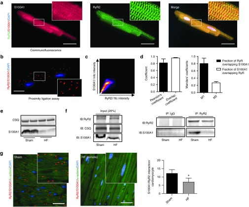
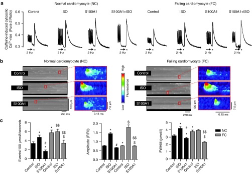
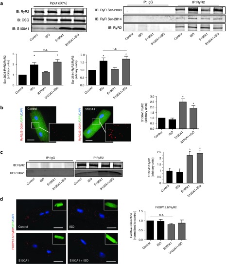
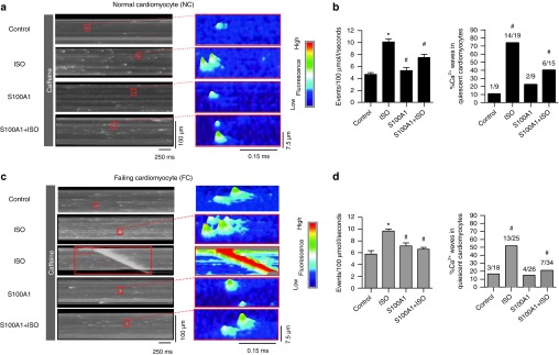
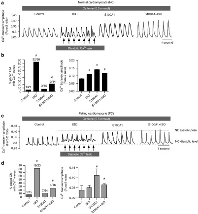
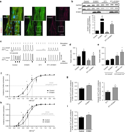
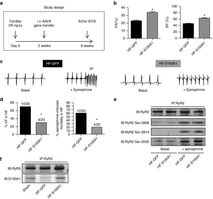
Restoring expression levels of the EF-hand calcium (Ca(2+)) sensor protein S100A1 has emerged as a key factor in reconstituting normal Ca(2+) handling in failing myocardium. Improved sarcoplasmic reticulum (SR) function with enhanced Ca(2+) resequestration appears critical for S100A1's cyclic adenosine monophosphate-independent inotropic effects but raises concerns about potential diastolic SR Ca(2+) leakage that might trigger fatal arrhythmias. This study shows for the first time a diminished interaction between S100A1 and ryanodine receptors (RyR2s) in experimental HF. Restoring this link in failing cardiomyocytes, engineered heart tissue and mouse hearts, respectively, by means of adenoviral and adeno-associated viral S100A1 cDNA delivery normalizes diastolic RyR2 function and protects against Ca(2+)- and β-adrenergic receptor-triggered proarrhythmogenic SR Ca(2+) leakage in vitro and in vivo. S100A1 inhibits diastolic SR Ca(2+) leakage despite aberrant RyR2 phosphorylation via protein kinase A and calmodulin-dependent kinase II and stoichiometry with accessory modulators such as calmodulin, FKBP12.6 or sorcin. Our findings demonstrate that S100A1 is a regulator of diastolic RyR2 activity and beneficially modulates diastolic RyR2 dysfunction. S100A1 interaction with the RyR2 is sufficient to protect against basal and catecholamine-triggered arrhythmic SR Ca(2+) leak in HF, combining antiarrhythmic potency with chronic inotropic actions.


Figures

Figure 1
Figure 1
Diminished interaction between S100A1 and RyR2 in postischemic heart failure. (a) Representative immunofluorescent images for S100A1 (red) and RyR2 (green) in a normal left ventricular rat cardiomyocyte reveal partial colocalization of S100A1 with RyR2 (merged image, yellow). Inlet magnification is threefold. (b) Representative immunofluorescent PLA image depicts in situ S100A1/RyR2 association (red dots) in a left ventricular cardiomyocyte. Each dot represents a complex consisting of S100A1 and RyR2. Inlet magnification is twofold. (c) Representative scatterplot image from colocalization analysis of Figure 1a using the JACoP plugin from ImageJ. (d) Colocalization analysis from RyR2/S100A1, depicting Person's, Overlap and Mander's coefficients, n = 3. (e) Representative immunoblots show diminished S100A1 protein abundance in postischemic HF mouse hearts versus sham. CSQ served as loading control. (f) Left: Representative input immunoblot for RyR2 immunoprecipitation from sham and HF mouse hearts. Right: Representative immunoblots of immunoprecipitated RyR2 and coprecipitating S100A1 protein from sham and postischemic HF mouse hearts. (g) Comparative in vivo PLA for S100A1 and RyR2 in sham (left) and HF (right) murine myocardium. Cardiomyocytes were counterstained with α-actinin (green). Inlet magnification is twofold, n = 3. *P < 0.05 versus sham. Data are given as mean ± SEM. Cardiomyocyte nuclei were counterstained with 4',6-diamidino-2-phenylindole (DAPI; blue). Scale bar represents 20 μm.
Figure 2
Figure 2
S100A1 attenuates physiological and abrogates pathological diastolic RyR2 activity. (a) Representative tracings of electrically-stimulated (2 Hz; arrow) steady-state Ca2+ transients from basal and ISO-stimulated (100 nmol/l) control (AdGFP) and S100A1-treated (AdS100A1) NCs (left) and FCs (right). Asterisk (*) indicates the 10 mmol/l caffeine pulse for semiquantitative assessment of the SR Ca2+ content due to the caffeine-mediated maximal rise of the cytosolic Ca2+ signal. Note that both ISO and S100A1 enhance the SR Ca2+ load in NCs and FCs (see Supplementary Figure S1d for statistical analyses). (b) Representative fluorescent line-scan and magnified surface plot images of elementary Ca2+ release events in control (AdGFP-transfected), isoproterenol-stimulated (ISO, 100 nmol/l, AdGFP-transfected) and S100A1 (AdS100A1-transfected) resting normal (NCs, left panel) and failing rat cardiomyocytes (FCs, right panel). (c) Corresponding statistical analysis of Ca2+ spark characteristics (frequency, amplitude, and width) shows that FCs exhibit significantly greater SR Ca2+ leak than NCs. S100A1 attenuates RyR2-mediated Ca2+ leak in NCs and FCs. n = 90 cells in each group derived from four different isolations. *P < 0.05 versus NC control, #P < 0.05 versus NC ISO, $P < 0.05 versus FC control, $$P < 0.05 versus FC ISO. Data are given as mean ± SEM.
Figure 3
Figure 3
Enhanced S100A1/RyR2 binding does not alter RyR2 phosphorylation and interaction with accessory proteins. (a) Left: Representative input immunoblot for RyR2 immunoprecipitation from control and S100A1-treated NCs. Right: Representative immunoblot of immunoprecipitated RyR2 and corresponding immunoblot for total RyR2, Ser-2808, and Ser-2814 phosphorylation from basal and ISO-stimulated (100 nmol/l) control (AdGFP) and corresponding S100A1-expressing (AdS100A1) cardiomyocytes (n = 5). Quantification is shown below. (b) Representative PLA image from control (AdGFP) and S100A1-treated (AdS100A1) rat cardiomyocytes showing S100A1/RyR2 interaction (red dots). Inlet magnification is twofold. Statistical analysis shows a twofold increase of the normalized arbitrary S100A1/RyR2 binding ratio (n = 4). (c) Representative immunoblot of immunoprecipitated RyR2 from basal and ISO-stimulated (100 nmol/l) control (AdGFP) and corresponding S100A1-expressing (AdS100A1) cardiomyocytes reveals a significant increase in coprecipitating S100A1 protein assessed by immunoblotting. Note that semiquantitative analysis of the normalized S100A1/RyR2 ratio matches results obtained with PLA (n = 5). (d) Representative PLA images for FKBP12.6/RyR2 interaction (red dots) in indicated groups. Inlets show the corresponding GFP-positive cardiomyocytes. Note that enhanced S100A1/RyR2 association did not change FKBP12.6 binding to the RyR2 (n = 4). Similar results were obtained for CaM and sorcin (Supplementary Figure S3b,c). *P < 0.05 versus control. Data are given as mean ± SEM. Nuclei (blue) were counterstained with 4',6-diamidino-2-phenylindole (DAPI). Scale bar represents 20 µm.
Figure 4
Figure 4
S100A1 prevents compound Ca2+ sparks and Ca2+ waves in quiescent ventricular cardiomyocytes with Ca2+ sensitized RyR2s. (a) and (c) Representative fluorescent line-scan and surface plot images of Ca2+ sparks in basal and ISO-stimulated (100 nmol/l) control (AdGFP) and S100A1-treated (AdS100A1) NCs (a) and FCs (c) with RyR2 sensitized to Ca2+ by low-dose (0.5 mmol/l) caffeine treatment. ISO triggers compound sparks and Ca2+ waves while S100A1-treatment entails solitary SR Ca2+ release events. Note that S100A1 inhibits the detrimental effects of ISO on SR Ca2+ leak. (b) and (d) Corresponding statistical analysis of Ca2+ spark frequency and Ca2+ wave incidence in NCs (b) and FCs (d) in indicated groups. n = 40 cells derived from four different isolations. #P < 0.05 versus ISO. Data are given as mean ± SEM.
Figure 5
Figure 5
S100A1 prevents premature diastolic Ca2+ waves during EC coupling in electro-mechanically coupled ventricular cardiomyocytes with “leaky” RyR2. (a) and (c) Representative tracings of Ca2+ transients of electrically-paced and to Ca2+ sensitized RyR2s basal and ISO-stimulated (100 nmol/l) control (AdGFP) and S100A1-treated (AdS100A1) NCs (left) and FCs (right). (b) and (d) Statistical analysis of diastolic Ca2+ waves and systolic Ca2+ transient amplitudes in electrically paced NCs (b) and FCs (d). Number of cells analyzed per group is shown on top of each bar. #P < 0.05 versus ISO. Data are given as mean ± SEM.
Figure 6
Figure 6
S100A1 restores isometric twitch tension and prevents premature diastolic after-contractions in engineered heart tissue (EHT). (a) Representative immunofluorescent staining displays a striated pattern of S100A1 (red) in cardiomyocytes in EHT. Cardiomyocytes were counterstained with actinin (green) and 4',6-diamidino-2-phenylindole (DAPI) (nuclei, blue). Scale bar represents 20 μm. Inlet magnification is threefold. (b) Representative immunoblots for GFP and S100A1 from normal and ET-1 stimulated (ET-1, 40 nmol/l, 96 hours stimulation) control (AdGFP) or S100A1-treated (AdS100A1) EHTs. Quantification of S100A1 protein levels normalized to GAPDH is shown below. n = 7, *P < 0.05 versus control, #P < 0.05 versus ET-1. (c) Representative electrically-stimulated isometric twitches from control- and S100A1-treated normal and HF-like EHT at 0.4 and 1.6 mmol/l extracellular Ca2+ ([Ca2+]extra). Lines indicate stimulation pulses (110 mA, 5 ms width, 2 Hz) inducing regular twitch; arrows indicate non-stimulated premature diastolic after-contractions. (d) Statistical analysis of twitch tension at 1.6 mmol/l [Ca2+]extra. n = 19 for control and S100A1, n = 17 for ET-1 and ET-1+S100A1, *P < 0.05 versus control, #P < 0.05 versus ET-1. (e) Statistical analysis of twitch tension at 0.2 mmol/l and 0.6 [Ca2+]extra/1 µmol/l ISO. n = 11, *P < 0.05 versus control. (f) Statistical analysis of premature diastolic after-contractions (in % of total twitches for each [Ca2+]extra) in control and S100A1-treated EHT. n = 15 for control and S100A1, *P < 0.05 versus control. (g) Calculated EC 50% ([Ca2+]extra at which 50% of EHTs shows regular after-contractions) in normal and ET-1 stimulated control and S100A1-treated EHT. n = 15 for control and S100A1, n = 8 for ET-1 and ET-1+S100A1; *P < 0.05 versus control/ET-1. (h) Statistical analysis of premature diastolic after-contractions (in % of total twitches for each [Ca2+]extra) in control and S100A1-treated EHT after βAR stimulation. n = 11, *P < 0.05 versus control. (i) Calculated EC 50% ([Ca2+]extra at which 50% of EHTs shows regular after-contractions) in normal and S100A1 transfected EHT after βAR stimulation. n = 11, *P < 0.05 versus control. Data are given as mean ± SEM.
Figure 7
Figure 7
S100A1 protects against epinephrine-triggered ventricular tachyarrhythmias and death in postischemic failing mouse hearts in vivo. (a) Study protocol following cardiac ischemia-reperfusion (I/R) damage due to temporary LAD occlusion. (b) Quantification of basal contractile performance assessed by echocardiography in anesthetized mice (FS, fractional shortening; EF, ejection fraction) of HF-GFP and HF-S100A1 mice at 6 weeks. n = 14, *P < 0.05 versus HF-GFP. (c) Representative telemetric ECG tracings of conscious HF mice (HF-GFP and HF-S100A1) depicting sinus rhythm before and development of ventricular fibrillation (VF) after i.p. injection of epinephrine (2 mg/kg body weight) in a HF-GFP mouse at week 6 of the study protocol. (d) Quantification of ventricular fibrillation (VF) and epinephrine-induced cardiac mortality in HF-GFP and HF-S100A1 mice. n = 20; *P < 0.05 versus HF-GFP. (e) Representative phospho-specific immunoblots for Ser-2808, Ser-2814, and Ser-20130 sites of immunoprecipated RyR2 from indicated groups under basal conditions and 20 minutes after epinephrine treatment. The experiment was repeated four times with identical results. (f) Representative immunoblots of immunoprecipitated RyR2 and coprecipitating S100A1 protein from sham, HF-GFP, and HF-S100A1 murine myocardium at week 6 of the study protocol. Data are given as mean ± SEM.

Similar articles

Cited by

References

    1. Bers, DM (2002). Cardiac excitation-contraction coupling. Nature 415: 198–205. - PubMed
    1. Bers, DM (2014). Cardiac sarcoplasmic reticulum calcium leak: basis and roles in cardiac dysfunction. Annu Rev Physiol 76: 107–127. - PubMed
    1. Morgan, JP, Erny, RE, Allen, PD, Grossman, W and Gwathmey, JK (1990). Abnormal intracellular calcium handling, a major cause of systolic and diastolic dysfunction in ventricular myocardium from patients with heart failure. Circulation 81(2 Suppl): III21–III32. - PubMed
    1. Farr, MA and Basson, CT (2004). Sparking the failing heart. N Engl J Med 351: 185–187. - PubMed
    1. Katz, AM (1986). Potential deleterious effects of inotropic agents in the therapy of chronic heart failure. Circulation 73(3 Pt 2): III184–III190. - PubMed

Publication types

MeSH terms